. 2023 Dec 20;6(1):115-125.
doi: 10.1007/s42995-023-00211-z. eCollection 2024 Feb.

A chitosan-based antibacterial hydrogel with injectable and self-healing capabilities

Affiliations

A chitosan-based antibacterial hydrogel with injectable and self-healing capabilities

Rui Chen et al. Mar Life Sci Technol. .

Abstract

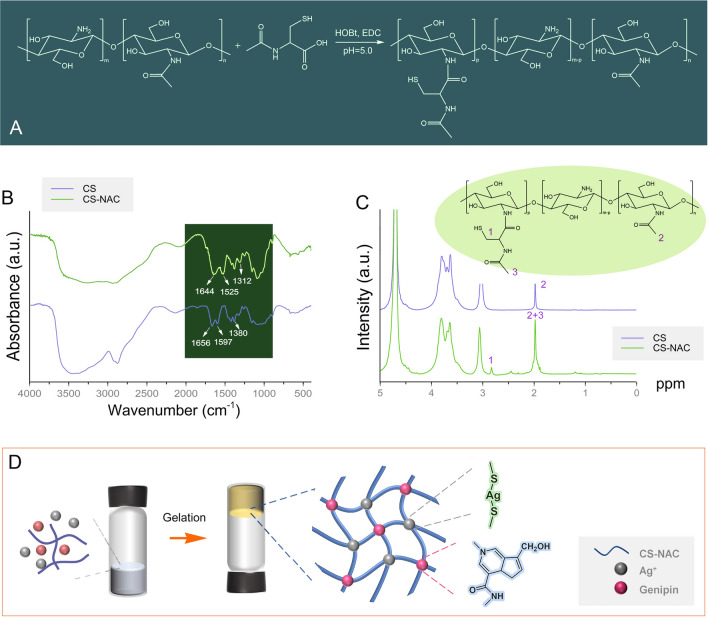
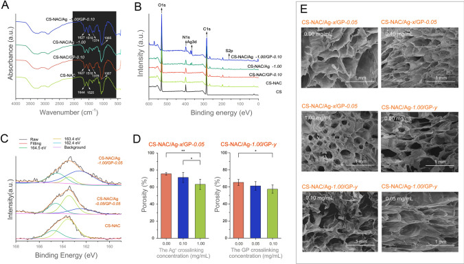
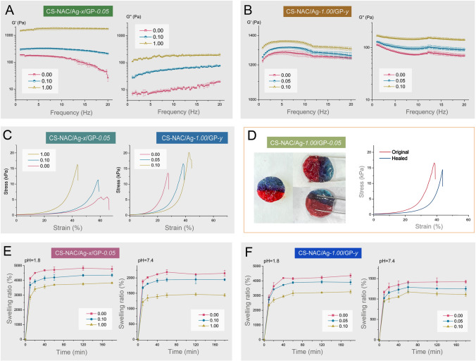
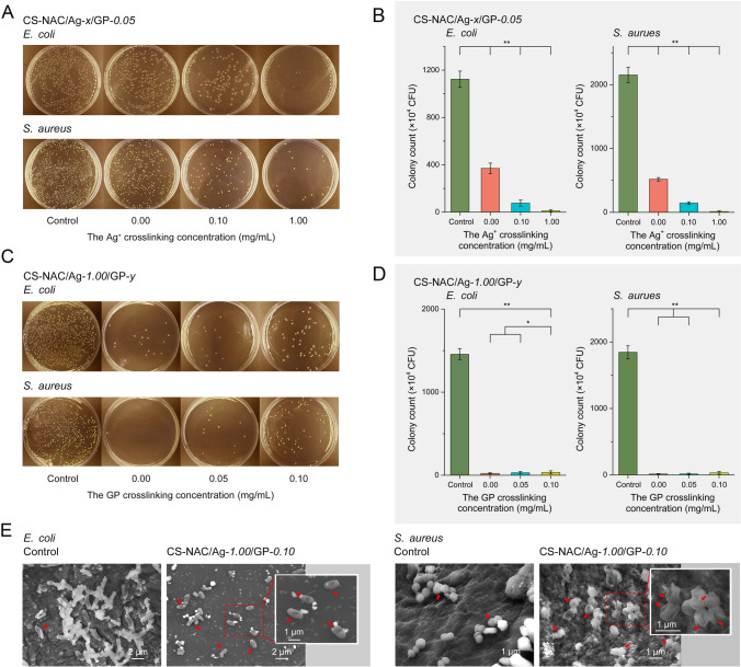
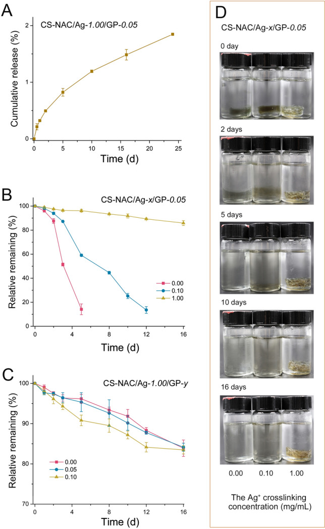
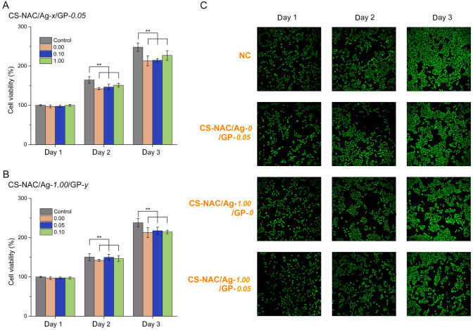
The presence of bacteria directly affects wound healing. Chitosan-based hydrogel biomaterials are a solution as they offer advantages for wound-healing applications due to their strong antimicrobial properties. Here, a double-cross-linking chitosan-based hydrogel with antibacterial, self-healing, and injectable properties is reported. Thiolated chitosan was successfully prepared, and the thiolated chitosan molecules were cross-linked by Ag-S coordination to form a supramolecular hydrogel. Subsequently, the amine groups in the thiolated chitosan covalently cross-linked with genipin to further promote hydrogel formation. In vitro experimental results indicate that hydrogel can release Ag+ over an extended time, achieving an antibacterial rate of over 99% against Escherichia coli and Staphylococcus aureus. Due to the reversible and dynamic feature of Ag-S coordination, an antibacterial hydrogel exhibited injectable and self-healing capabilities. Additionally, the hydrogel showed excellent biocompatibility and biodegradability.

Supplementary information: The online version contains supplementary material available at 10.1007/s42995-023-00211-z.

Keywords: Antibacterial; Chitosan; Cross-linking; Hydrogel; Silver ion.


Conflict of interest statement

Conflict of interestThe authors declare they have no conflict of interest.

Figures

Fig. 1
Fig. 1
A Synthesis of CS-NAC; B FT-IR spectra of CS-NAC and chitosan; C 1H NMR spectra of CS-NAC and chitosan; and D schematic architecture of CS-NAC/Ag+/GP hydrogels
Fig. 2
Fig. 2
Characterization of CS-NAC/Ag+/GP hydrogels with different Ag+ and genipin concentrations. A FT-IR spectra, B XPS patterns, C XPS S2p spectra, D porosity, and E SEM images of the hydrogels (n = 3, mean ± SD, *P < 0.05 and **P < 0.01)
Fig. 3
Fig. 3
A Frequency sweeps (G′ and G″) of the CS-NAC/Ag+/GP hydrogels with different Ag+ concentrations (1 mm gap, n = 3, mean ± SD); B frequency sweeps of the hydrogels with various concentrations of genipin (n = 3, mean ± SD); C the compressive stress–strain curves of the hydrogels with different concentrations of Ag+ and genipin; D photographs of healing processes and compressive stress–strain curves of before and after wound healing; and E swelling ratio of the CS-NAC/Ag+/GP hydrogels (n = 3, mean ± SD)
Fig. 4
Fig. 4
In vitro antibacterial activity of the hydrogels cross-linked with various Ag+ or genipin concentrations against E. coli and S. aureus. A and C Photographs of agar plates and B and D corresponding statistical data of the colonies of E. coli and S. aureus. (n = 6, mean ± SD, *P < 0.05 and **P < 0.01). E SEM images showing the morphological changes of E. coli and S. aureus after incubation of the CS-NAC/Ag+-1.00/GP-0.05 hydrogel
Fig. 5
Fig. 5
A Cumulative release profiles of Ag+ from the CS-NAC/Ag+-1.00/GP-0.05 hydrogel (n = 3, mean ± SD). B Degradation behavior of the hydrogels with different Ag+ concentrations (n = 4, mean ± SD). C Degradation behavior of the hydrogels with various concentrations of GP. D Photographs of the degraded hydrogels
Fig. 6
Fig. 6
Biocompatibility analysis. A, B Cell viability of fibroblast cells cultured in the hydrogel-conditioned media. C Live/dead staining fluorescent images (n = 4, mean ± SD, *P < 0.05 and **P < 0.01)

References

    1. Alshameri AW, Owais M. Antibacterial and cytotoxic potency of the plant-mediated synthesis of metallic nanoparticles Ag NPs and ZnO NPs: a review. OpenNano. 2022;8:100077. doi: 10.1016/j.onano.2022.100077. - DOI
    1. Bapat RA, Chaubal TV, Joshi CP, Bapat PR, Choudhury H, Pandey M, Gorain B, Kesharwani P. An overview of application of silver nanoparticles for biomaterials in dentistry. Mater Sci Eng C. 2018;91:881–898. doi: 10.1016/j.msec.2018.05.069. - DOI - PubMed
    1. Chen K-Y, Liao W-J, Kuo S-M, Tsai F-J, Chen Y-S, Huang C-Y, Yao C-H. Asymmetric chitosan membrane containing collagen I nanospheres for skin tissue engineering. Biomacromol. 2009;10:1642–1649. doi: 10.1021/bm900238b. - DOI - PubMed
    1. Dai X, Guo Q, Zhao Y, Zhang P, Zhang T, Zhang X, Li C. Functional silver nanoparticle as a benign antimicrobial agent that eradicates antibiotic-resistant bacteria and promotes wound healing. ACS Appl Mater Interfaces. 2016;8:25798–25807. doi: 10.1021/acsami.6b09267. - DOI - PubMed
    1. Ellman GL. Tissues sulfhydryl groups. Arch Biochem Biophys. 1959;82:70–77. doi: 10.1016/0003-9861(59)90090-6. - DOI - PubMed

LinkOut - more resources