

Color translation from monoscopic photogrammetry +ID Methodology into a Polyjet final 3D printed facial prosthesis
- PMID: 38434006
- PMCID: PMC10904947
- DOI: 10.12688/f1000research.111196.1
Color translation from monoscopic photogrammetry +ID Methodology into a Polyjet final 3D printed facial prosthesis
Abstract
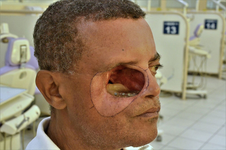
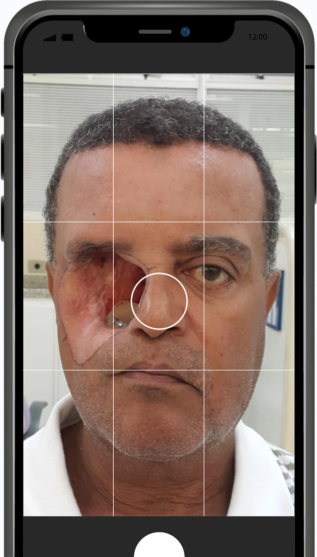
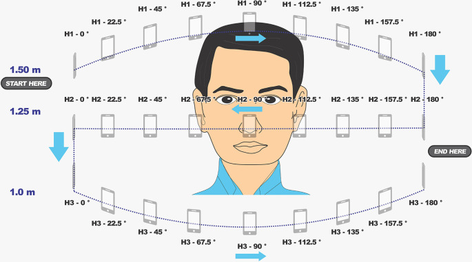
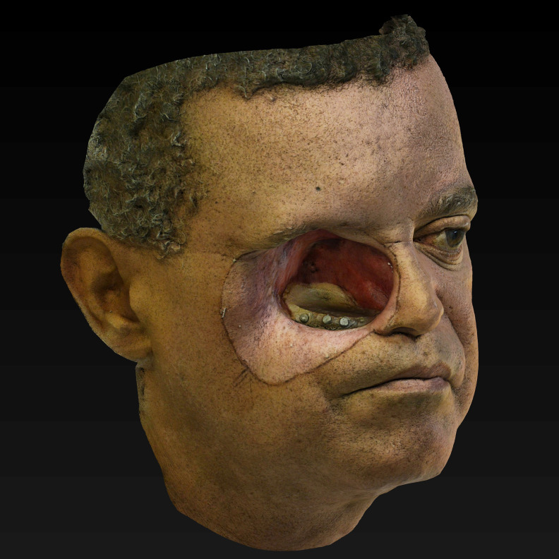
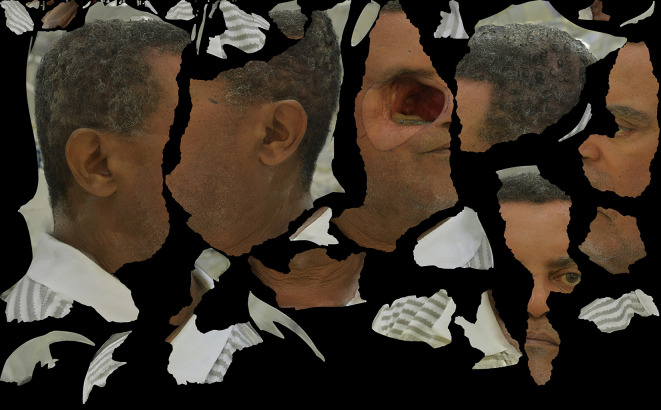
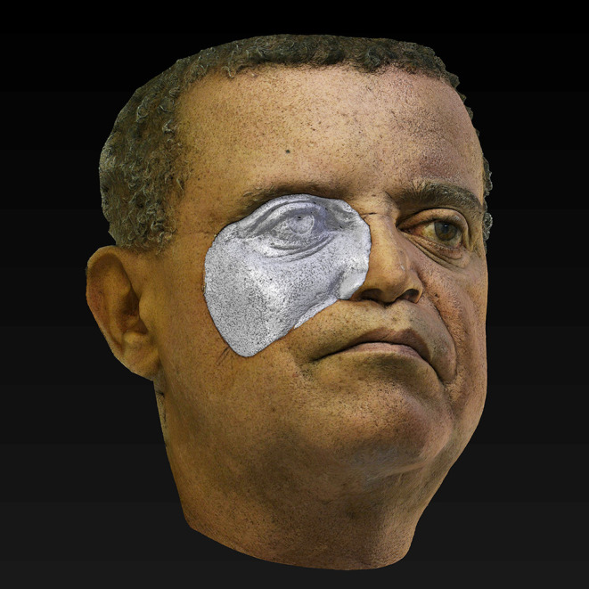
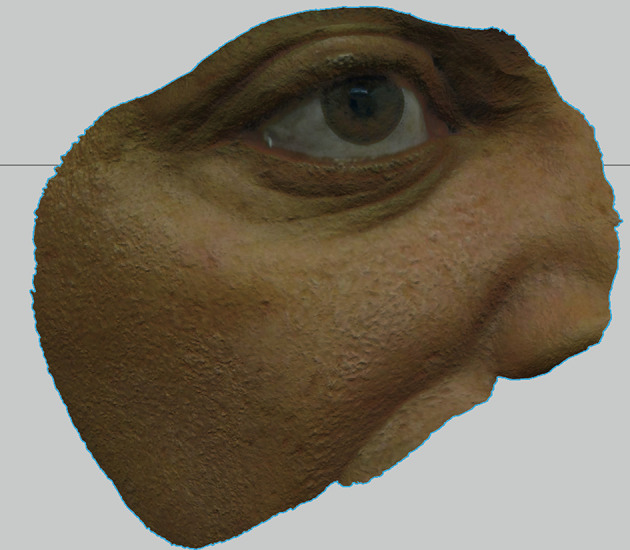
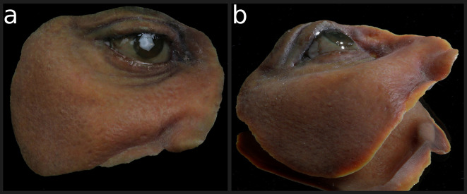
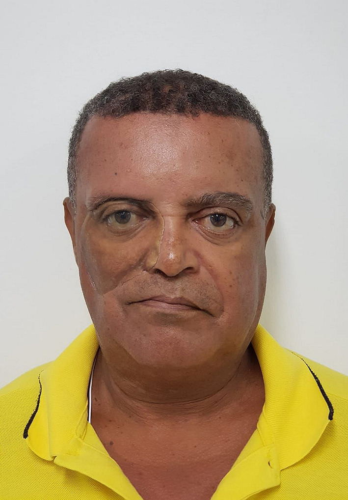
Background: The artistic techniques necessary to fabricate facial prostheses mainly depend on individual skill and are not a resource easily reproduced. Digital technology has contributed to improved outcomes, often combining analog and new digital techniques in the same workflow. Methods: This article aims to present an innovative workflow to produce a final colored 3D printed and facial prosthesis by UV-map color translation into colored resin 3D printing. A modified +ID Methodology was used to obtain 3D models with the calibrated 3D printable patient's skin color. No hands-on physical molding, manual sculpture, or intrinsic silicone coloration was used. Results: The outcome resulted in acceptable aesthetics, adaptation, and an approximate color match after extrinsic coloration. The patient reported good comfort and acceptance. Conclusions: A direct resin 3D printed prosthesis may be a viable alternative, especially for rapid delivery as an immediate prosthesis or an option when there is no experienced anaplastogist to manufacture a conventional prosthesis.
Keywords: 3D printing; color; head and neck neoplasms; maxillofacial prosthesis; prosthesis design.
Copyright: © 2022 Salazar-Gamarra R et al.
Conflict of interest statement
No competing interests were disclosed.
Figures
Similar articles
-
Evaluation of accuracy of photogrammetry with 3D scanning and conventional impression method for craniomaxillofacial defects using a software analysis.Trials. 2022 Dec 27;23(1):1048. doi: 10.1186/s13063-022-07005-1. Trials. 2022. PMID: 36575547 Free PMC article. Clinical Trial.
-
Direct 3D Printing of Flexible Nasal Prosthesis: Optimized Digital Workflow from Scan to Fit.J Prosthodont. 2019 Jan;28(1):10-14. doi: 10.1111/jopr.13001. Epub 2018 Nov 29. J Prosthodont. 2019. PMID: 30461125
-
[Application of negative molds technology based on three-dimensional printing in digital maxillofacial prostheses].Zhonghua Kou Qiang Yi Xue Za Zhi. 2017 Jun 9;52(6):336-341. doi: 10.3760/cma.j.issn.1002-0098.2017.06.003. Zhonghua Kou Qiang Yi Xue Za Zhi. 2017. PMID: 28613053 Chinese.
-
A systematic review of the computerized tools and digital techniques applied to fabricate nasal, auricular, orbital and ocular prostheses for facial defect rehabilitation.J Stomatol Oral Maxillofac Surg. 2020 Jun;121(3):268-277. doi: 10.1016/j.jormas.2019.10.003. Epub 2019 Oct 11. J Stomatol Oral Maxillofac Surg. 2020. PMID: 31610244
-
A shade guide for acrylic resin facial prostheses.J Prosthet Dent. 1992 Jul;68(1):120-2. doi: 10.1016/0022-3913(92)90299-p. J Prosthet Dent. 1992. PMID: 1403901 Review.
Cited by
-
Present and future of extraoral maxillofacial prosthodontics: Cancer rehabilitation.Front Oral Health. 2022 Oct 19;3:1003430. doi: 10.3389/froh.2022.1003430. eCollection 2022. Front Oral Health. 2022. PMID: 36338571 Free PMC article. Review.
References
-
- Farook TH, Jamayet NB, Abdullah JY, et al. : A systematic review of the computerized tools and digital techniques applied to fabricate nasal, auricular, orbital and ocular prostheses for facial defect rehabilitation. J. Stomatol. Oral Maxillofac. Surg. 2019;121:268–277. 10.1016/j.jormas.2019.10.003 - DOI - PubMed
-
- Salazar-Gamarra R, et al. : Introdução à Metodologia Mais Identidade Proteses Faciais 3D com a utilização de tecnologias accessíveis para pacientes sobreviventes de cancer no rosto. E-book Comunicação Científica eTécnica em Odontologia. 2019;251–272. 10.22533/at.ed.26519290320 - DOI
Associated data
LinkOut - more resources
Full Text Sources