

Mitotherapy inhibits against tenofovir induced nephrotoxicity on rat renal proximal tubular cells
- PMID: 38434141
- PMCID: PMC10907186
- DOI: 10.1016/j.bbrep.2024.101669
Mitotherapy inhibits against tenofovir induced nephrotoxicity on rat renal proximal tubular cells
Abstract
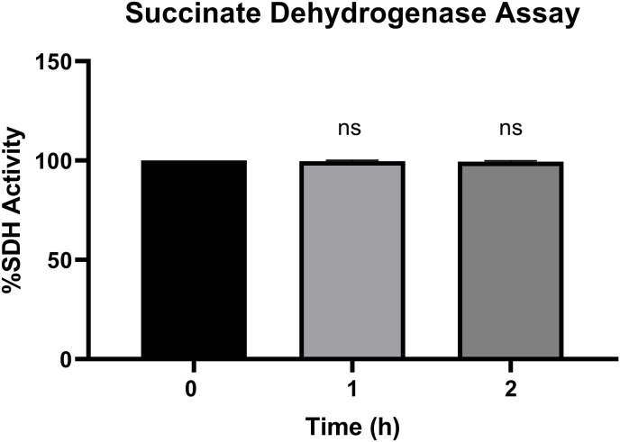
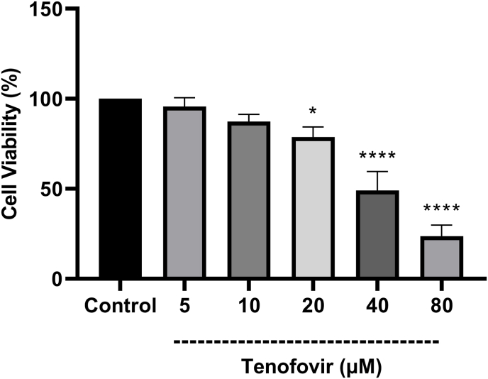
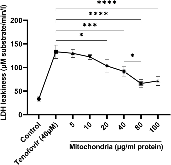
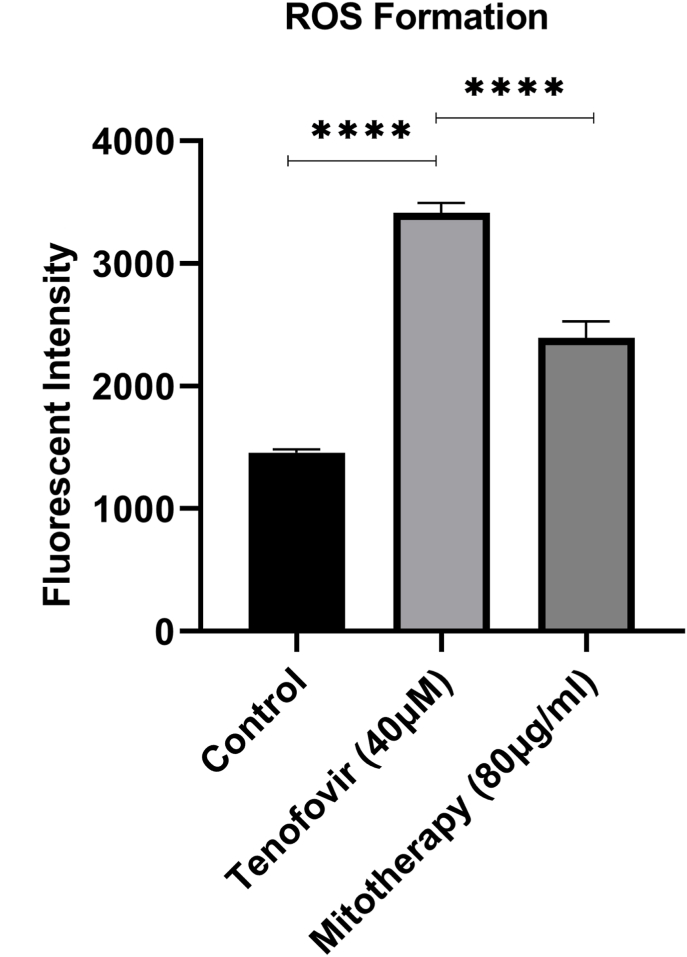
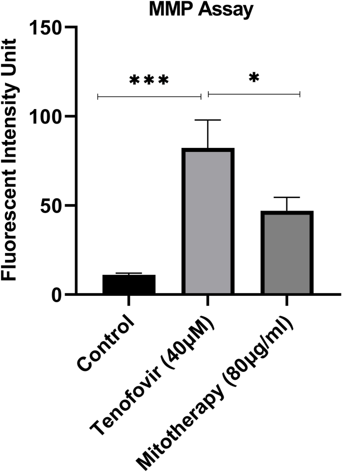
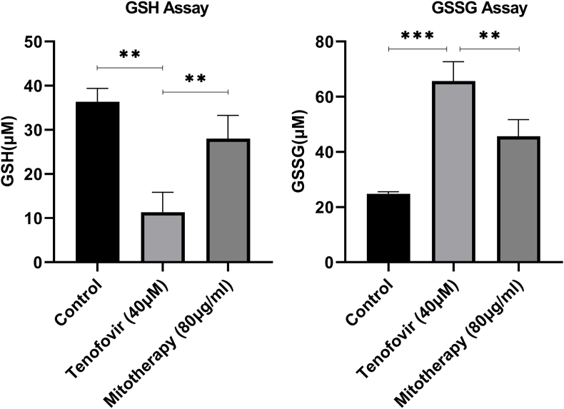
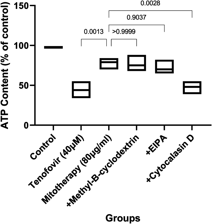
Tenofovir, as nucleotide reverse transcriptase inhibitors (NRTIs), is used to prevent and cure HIV/AIDS. Ample evidence confirmed that the nephrotoxicity of tenofovir has been linked to mitochondrial dysfunction. It seems that transplantation with healthy mitochondria instead of damaged mitochondria may be a beneficial approach to therapy. Therefore, it decided to investigate the impact of mitotherapy on tenofovir against renal proximal tubular cells (RPTCs) toxicity by measurement of oxidative stress and cytotoxicity biomarkers and restoring of mitochondrial function on isolated mitochondria. EC50 of tenofovir was achieved at 40 μM following 2 h incubation in Earle's solution (pH = 7.4; 37 °C). Freshly isolated mitochondria (80 μg/ml) were added to damage RPTCs affected by tenofovir in treated groups. One Way ANOVA analysis showed that healthy mitochondrial transplantation decreased oxidative stress biomarkers following tenofovir toxicity in RPTCs. Our data revealed that mitotherapy makes cell survival possible in RPTCs affected by tenofovir. In addition, it supposed that a novel and ideal strategy for the treatment of chemicals-induced nephrotoxicity.
Keywords: Mitochondrial transplantation; Nephrotoxicity; Oxidative stress; Renal proximal tubular cells (RPTCs); Tenofovir.
© 2024 The Authors.
Conflict of interest statement
The authors declare that they have no known competing financial interests or personal relationships that could have appeared to influence the work reported in this paper.
Figures
References
-
- De Clercq E. In search of a selective therapy of viral infections. Antivir. Res. 2010;85(1):19–24. - PubMed
-
- Dietz J.-P., et al. Di-tert-butyl phosphonate route to the antiviral drug tenofovir. Org. Process Res. Dev. 2021;25(4):789–798. - PubMed
-
- Zimmermann A.E., et al. Tenofovir-associated acute and chronic kidney disease: a case of multiple drug interactions. Clin. Infect. Dis. 2006;42(2):283–290. - PubMed
-
- Lewis W., Copeland W.C., Day B.J. Mitochondrial DNA depletion, oxidative stress, and mutation: mechanisms 0f dysfunction from nucleoside reverse transcriptase inhibitors. Lab. Invest. 2001;81(6):777–790. - PubMed
-
- Lewis W. Nucleoside reverse transcriptase inhibitors, mitochondrial DNA and AIDS therapy. Antivir. Ther. 2005;10(2_suppl):13–27. - PubMed
LinkOut - more resources
Full Text Sources
