Case Reports
. 2024 Feb 16:15:1339438.
doi: 10.3389/fneur.2024.1339438. eCollection 2024.

Case report: Susac syndrome-two ends of the spectrum, single center case reports and review of the literature

Affiliations
Case Reports

Case report: Susac syndrome-two ends of the spectrum, single center case reports and review of the literature

Martina Cviková et al. Front Neurol. .

Abstract

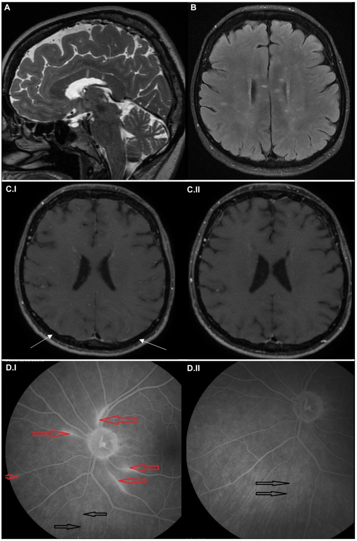
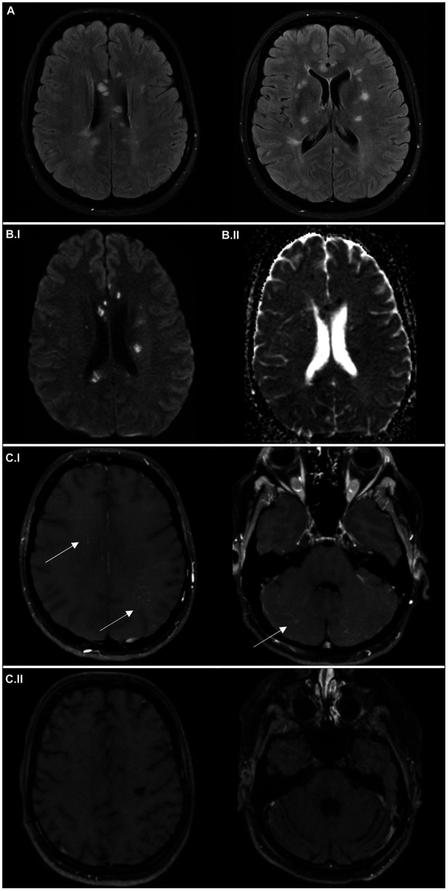
Susac syndrome is a rare and enigmatic complex neurological disorder primarily affecting small blood vessels in the brain, retina, and inner ear. Diagnosing Susac syndrome may be extremely challenging not only due to its rarity, but also due to the variability of its clinical presentation. This paper describes two vastly different cases-one with mild symptoms and good response to therapy, the other with severe, complicated course, relapses and long-term sequelae despite multiple therapeutic interventions. Building upon the available guidelines, we highlight the utility of black blood MRI in this disease and provide a comprehensive review of available clinical experience in clinical presentation, diagnosis and therapy of this disease. Despite its rarity, the awareness of Susac syndrome may be of uttermost importance since it ultimately is a treatable condition. If diagnosed in a timely manner, early intervention can substantially improve the outcomes of our patients.

Keywords: Susac syndrome; black blood imaging; branch retinal arterial occlusion; hearing loss; neuroimmunology; stroke; vasculitis.


Conflict of interest statement

The authors declare that the research was conducted in the absence of any commercial or financial relationships that could be construed as a potential conflict of interest.

Figures

Figure 1
Figure 1
Imaging findings in the case report 1. Sagittal T2-weighted (A) and axial FLAIR MRI scans (B) at the time of symptom onset showing multiple hyperintense small lesions in the white matter, predominantly affecting corpus callosum in the form of “spokes.” Axial black blood MRI scans at the time of symptom onset (C.I), showing microangiopathy as multiple small, hypersignal nodules in small vessel walls combined with discreet leptomemingeal enhancement (marked with arrows); and after 6 months (C.II), showing regression of the pathology. Fluorescein angiogram (left eye) at the time of symptom onset (D.I), where the areas of hyperfluorescence mark leakage from inflamed retinal arterioles (red arrows) and the area of fluorescence decrease corresponds to the lower nasal arteriolar occlusion (precapillary plaque) with segmentation (black arrows). Fluorescein angiogram (left eye) after 8 months (D.II) shows the absence of leakage and reperfusion of the previously occluded retinal arterioles (black arrows).
Figure 2
Figure 2
Imaging findings in the case report 2. Axial FLAIR MRI scans (A) at the time of symptom onset showing multiple hyperintense “snowball” lesions in the white matter. Axial average diffusion-weighted scans (B.I) at the time of symptom onset and apparent diffusion coefficient maps (B.II), with small areas of restriction of diffusion (hypersignal in B.I and hyposignal in B.II), corresponding to small infarctions. Black blood MRI scans at the time of symptom onset (C.I), detecting microangiopathy (multiple small hypersignal nodules of small vessel walls) in both supra- and infratentorial region; and after 6 months (C.II), with regression of the initial pathology, but development of substantial atrophy.
Figure 3
Figure 3
Imaging findings in the case report 2. Axial FLAIR MRI scans (A) about 1.5 months after the symptom onset, showing cystoid degeneration of previous hypersignal areas and several new lesions. Axial average diffusion-weighted scans (B.I) and apparent diffusion coefficient maps (B.II) about 1.5 months after the symptom onset, with several new lesions causing diffusion restriction, and 6 months after the symptom onset (C.I,C.II), where no new lesions are apparent.
Figure 4
Figure 4
Imaging findings in the case report 2 – Fluorescein angiogram of the left (A.I) and right eye (A.II) 1.5 months after symptom onset, where the areas of hyperfluorescence mark leakage from inflamed retinal arterioles (red arrow) and the areas of fluorescence decrease correspond to arteriolar occlusion (precapillary plaque) (black arrows). Follow-up fluorescein angiogram (left eye) after 6 months (B) showing revascularization of small vessel occlusions present in the previous examination (black arrow); no areas of hyperfluorescence were detected.

Similar articles

References

    1. Dörr J, Krautwald S, Wildemann B, Jarius S, Ringelstein M, Duning T, et al. . Characteristics of Susac syndrome: a review of all reported cases. Nat Rev Neurol. (2013) 9:307–16. doi: 10.1038/nrneurol.2013.82, PMID: - DOI - PubMed
    1. Zengin Karahan S, Boz C, Saip S, Kale N, Demirkaya S, Celik Y, et al. . Susac syndrome: clinical characteristics, diagnostic findings and treatment in 19 cases. Mult Scler Relat Disord. (2019) 33:94. doi: 10.1016/j.msard.2019.05.018 - DOI - PubMed
    1. Triplett JD, Qiu J, O'Brien B, Gopinath S, Trewin B, Spring PJ, et al. . Diagnosis, differential diagnosis and misdiagnosis of Susac syndrome. Eur J Neurol. (2022) 29:1771–81. doi: 10.1111/ene.15317, PMID: - DOI - PMC - PubMed
    1. David C, Sacré K, Henri-Feugeas MC, Klein I, Doan S, Cohen FA, et al. . Susac syndrome: a scoping review. Autoimmun Rev. (2022) 21:103097. doi: 10.1016/j.autrev.2022.103097, PMID: - DOI - PubMed
    1. Dekeyser C, Vanhoorne A, Hemelsoet D, van Hijfte L, de Zaeytijd J, van Driessche V, et al. . Atypical clinical and novel radiological findings in Susac syndrome: experience from a large monocentric cohort. J Neuroimmunol. (2023) 376:578032. doi: 10.1016/j.jneuroim.2023.578032, PMID: - DOI - PubMed

Publication types