Isolated right ventricular hypoplasia associated with cyanotic atrial septal defect: a case report
- PMID: 38434213
- PMCID: PMC10908390
- DOI: 10.1093/ehjcr/ytae094
Isolated right ventricular hypoplasia associated with cyanotic atrial septal defect: a case report
Abstract
Background: Hypoxaemia in isolated right ventricular (RV) hypoplasia (IRVH) is primarily caused by a right-to-left shunt (RLS) at the atrial level, such as an atrial septal defect (ASD). When considering closure of the RLS, it should be closed only after ensuring that it will not cause right-sided heart failure (HF).
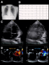
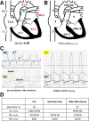
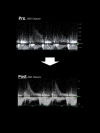
Case summary: A 21-year-old woman had been experiencing shortness of breath during exertion since childhood. Transthoracic and transoesophageal echocardiography revealed an ASD with bidirectional shunting, and microbubble test revealed a marked RLS. Cardiac magnetic resonance imaging revealed a hypoplastic RV end-diastolic volume corrected for body surface area of 47 mL/m2 (70% of normal range). Right heart catheterization revealed a decreased Qp/Qs ratio of 0.89 and a pressure waveform with a clear increase in the 'A'-wave, although the mean right atrial pressure was not high (4 mmHg). Therefore, the patient was diagnosed with cyanotic ASD and IRVH. A temporary balloon occlusion test was performed to evaluate the right-sided heart response to capacitive loading prior to ASD closure. After treatment, the patient's improved markedly. The pre-operative brain natriuretic peptide (BNP) level was normal; however, 6 months after ASD closure, the BNP level was elevated, and the continuous-wave Doppler waveform of pulmonary regurgitation at the time of transthoracic echocardiography changed, suggesting an increase in diastolic RV pressure.
Discussion: When ASD is complicated by hypoxaemia, the possibility of IRVH, although rare, should be considered. Another difficult point is determining whether the ASD can be closed, considering its immature RV compliance.
Keywords: Balloon occlusion test; Case report; Cyanotic atrial septal defect; Isolated right ventricular hypoplasia.
© The Author(s) 2024. Published by Oxford University Press on behalf of the European Society of Cardiology.
Conflict of interest statement
Conflict of interest: None declared.
Figures


Similar articles
-
Temporary atrial septal defect balloon occlusion test as a must in the elderly.BMC Cardiovasc Disord. 2023 Jan 12;23(1):15. doi: 10.1186/s12872-023-03046-9. BMC Cardiovasc Disord. 2023. PMID: 36635628 Free PMC article.
-
Transcatheter closure of atrial septal defect in adults: time-course of atrial and ventricular remodeling and effects on exercise capacity.Int J Cardiovasc Imaging. 2019 Nov;35(11):2077-2084. doi: 10.1007/s10554-019-01647-0. Epub 2019 Jun 15. Int J Cardiovasc Imaging. 2019. PMID: 31203534 Free PMC article.
-
Right Ventricle before and after Atrial Septal Defect Device Closure.Echocardiography. 2016 Sep;33(9):1381-8. doi: 10.1111/echo.13250. Epub 2016 Apr 24. Echocardiography. 2016. PMID: 27109837 Clinical Trial.
-
[A case report: successful direct closure of atrial septal defect in isolated right ventricular hypoplasia].Kyobu Geka. 1992 Feb;45(2):179-82. Kyobu Geka. 1992. PMID: 1542200 Review. Japanese.
-
Fenestrated Transcatheter ASD Closure in Adults with Diastolic Dysfunction and/or Pulmonary Hypertension: Case Series and Review of the Literature.Congenit Heart Dis. 2016 Dec;11(6):663-671. doi: 10.1111/chd.12367. Epub 2016 Apr 29. Congenit Heart Dis. 2016. PMID: 27125263 Review.
References
-
- Marrone G, Mamone G, Luca A, Vitulo P, Bertani A, Pilato M, et al. The role of 1.5 T cardiac MRI in the diagnosis, prognosis and management of pulmonary arterial hypertension. Int J Cardiovasc Imaging 2010;26:665–681. - PubMed
-
- Krasemann T, van Osch-Gevers L, van de Woestijne P. Cyanosis due to an isolated atrial septal defect: case report and review of the literature. Cardiol Young 2020;30:1741–1743. - PubMed
Publication types
LinkOut - more resources
Full Text Sources
Research Materials
Miscellaneous
