. 2023 Nov 22:6:0273.
doi: 10.34133/research.0273. eCollection 2023.

Cortical Organoid-on-a-Chip with Physiological Hypoxia for Investigating Tanshinone IIA-Induced Neural Differentiation

Affiliations

Cortical Organoid-on-a-Chip with Physiological Hypoxia for Investigating Tanshinone IIA-Induced Neural Differentiation

Yue Zhi et al. Research (Wash D C). .

Abstract

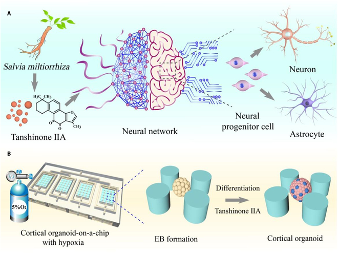
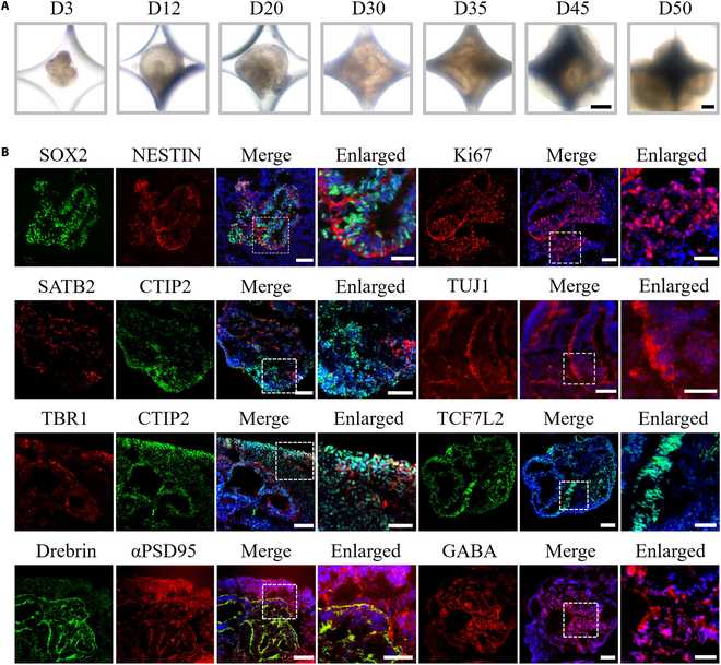
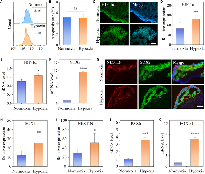
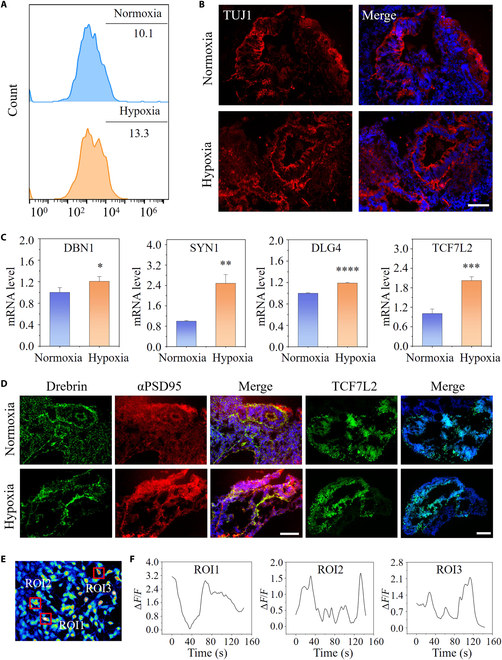
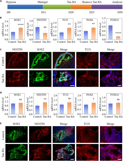
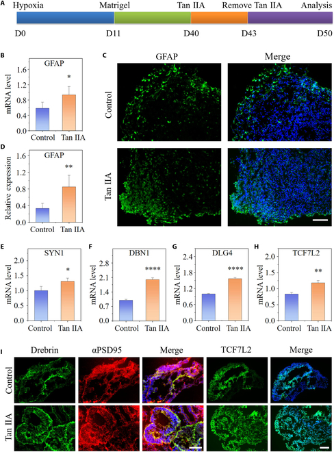
Cortical organoids represent cutting-edge models for mimic human brain development during the early and even middle stage of pregnancy, while they often fail to recreate the complex microenvironmental factors, such as physiological hypoxia. Herein, to recapitulate fetal brain development, we propose a novel cortical organoid-on-a-chip with physiological hypoxia and further explore the effects of tanshinone IIA (Tan IIA) in neural differentiation. The microfluidic chip was designed with a micropillar array for the controlled and efficient generation of cortical organoids. With low oxygen, the generated cortical organoids could recapitulate key aspects of early-gestational human brain development. Compared to organoids in normoxic culturing condition, the promoted neurogenesis, synaptogenesis and neuronal maturation were observed in the present microsystem, suggesting the significance of physiological hypoxia in cortical development. Based on this model, we have found that Chinese herbal drug Tan IIA could promote neural differentiation and maturation, indicating its potential therapeutic effects on neurodevelopmental disorders as well as congenital neuropsychiatric diseases. These results indicate that the proposed biomimetic cortical organoid-on-a-chip model with physiological hypoxia can offer a promising platform to simulate prenatal environment, explore brain development, and screen natural neuroactive components.


Conflict of interest statement

Competing interests: The authors declare that they have no competing interests.

Figures

Fig. 1.
Fig. 1.
Illustration of the microsystem with physiological hypoxia, allowing to probe the effects of Tan IIA in neurodevelopment. (A) Tan IIA promotes the formation and function of neural networks, which is reflected in the differentiation and maturation of various types of cells. (B) Scheme showing human cortical organoids exposed to low oxygen in a hypoxic incubator. The overall structure of the microsystem and the enlarged image of the internal details show the corticogenesis process.
Fig. 2.
Fig. 2.
Formation and validation of the human cortical organoid under hypoxic conditions. (A) Representative bright-field pictures of the cortical organoids generated on the chip within the hypoxic environment. Scale bars, 500 μm. (B) The immunofluorescence images of NPCs (SOX2 and NESTIN), proliferative marker Ki67, cortical layer (TBR1, SATB2, and CTIP2), differentiated neurons (TUJ1), postsynapse (αPSD95 and drebrin), Wnt signaling pathway (TCF7L2), and inhibitory neurons (GABA) within 40-d cortical organoids. Scale bars, 100 μm (in “Merge”) and 50 μm (in “Enlarged”).
Fig. 3.
Fig. 3.
Cellular responses to hypoxia in cortical organoids. (A and B) Fluorescence-activated cell sorting analysis and quantitative results of apoptosis in hypoxia- and normoxia-exposed cortical organoids at day 30 (n = 3). (C and D) Representative immunostaining and quantitative analysis of HIF-1α in cortical organoids exposed for 30 d to 5% O2 versus 21% O2 (n = 6). (E and F) Expression of HIF-1α and SOX2 at the mRNA level in cortical organoids under normoxic or hypoxic conditions at day 30. (G) Immunofluorescence images for SOX2- and NESTIN-positive areas in cortical organoids under low oxygen for 30 d. (H and I) The fluorescence intensity of SOX2 and NESTIN was quantified at day 30 (n = 6). (J and K) mRNA Expression of PAX6 and FOXG1 in cortical organoids under normoxia or hypoxia at day 30 was identified by qRT-PCR (n = 3). All data are the means of at least 3 replicates ± SD. The data were analyzed using the Student t test (ns, not significant; *P < 0.05; **P < 0.01; ***P < 0.001; ****P < 0.0001). Scale bars, 50 μm (C) and 100 μm (G).
Fig. 4.
Fig. 4.
Neuron differentiation and neural network formation under hypoxic conditions. (A) Fluorescence-activated cell sorting analysis of TUJ1+ in hypoxia- and normoxia-exposed cortical organoids at day 30. (B) TUJ1+ cells in cortical organoids treated with hypoxia or normoxia at day 30 were identified by immunohistochemical analysis. (C) The relative mRNA expression of synaptic relative genes at day 50 (SYN1, DBN1, DLG4, and TCF7L2) was determined by qRT-PCR (n = 3). (D) Immunostaining images of postsynaptic markers (drebrin and αPSD95) and Wnt-related marker (TCF7L2) with the treatment of hypoxia or normoxia at day 50 are shown. (E and F) Calcium imaging of hypoxia-treated cortical organoids at day 30 as assessed using fluorescence alterations. ΔF/F: relative concentration of Ca2+. Red boxes designated ROIs (regions of interest). All data are the means of at least 3 replicates ± SD. The data were analyzed using the Student t test (ns, not significant; *P < 0.05; **P < 0.01; ***P < 0.001; ****P < 0.0001). Scale bars, 100 μm.
Fig. 5.
Fig. 5.
Tan IIA-induced neuronal differentiation and corticogenesisin organoids under hypoxia. (A) Experimental flow chart of treatment of cortial organoid with Tan IIA at an earlier developmental stage. (B and D) Expression of neural genes at the mRNA level in cortical organoids with the presence or absence of Tan IIA at days 30 (B) and 50 (D) (n = 3). (C and E) Immunostaining images of SOX2, NESTIN, and TUJ1 with treatment of Tan IIA or not at days 30 (C) and 50 (E) are shown. All data are the means of at least 3 replicates ± SD. The data were analyzed using the Student t test (ns, not significant; *P < 0.05; **P < 0.01; ****P < 0.0001). Scale bars, 100 μm.
Fig. 6.
Fig. 6.
Tan IIA-induced functional differentiation and neural network maturation in cortical organoids under hypoxia. (A) Experimental flow chart of treatment of cortial organoid with Tan IIA at a later developmental stage. (B) Expression of GFAP by qRT-PCR within cortical organoids under the treatment of Tan IIA or not at day 50 (n = 3). (C and D) Immunofluorescent staining for GFAP and quantifications for the expression of GFAP in cortical organoids with and without Tan IIA treatment at day 50 (n = 6). (E to H) Comparison of relative mRNA expression in synaptic relative genes within 50-d cortical organoids with or without Tan IIA treatment by qRT-PCR (n = 3). (I) Immunohistochemical staining for αPSD95, drebrin, and TCF7L2 in cortical organoids at day 50. All data are the means of at least 3 replicates ± SD. The data were analyzed using the Student t test (*P < 0.05; **P < 0.01; ****P < 0.0001). Scale bars, 100 μm.

Similar articles

Cited by

References

    1. Juric-Sekhar G, Hevner RF. Malformations of cerebral cortex development: Molecules and mechanisms. Annu Rev Pathol. 2019;14:293–318. - PMC - PubMed
    1. Klingler E, Francis F, Jabaudon D, Cappello S. Mapping the molecular and cellular complexity of cortical malformations. Science. 2021;371(6527):eaba4517. - PubMed
    1. Paulsen B, Velasco S, Kedaigle AJ, Pigoni M, Quadrato G, Deo AJ, Adiconis X, Uzquiano A, Sartore R, Yang SM, et al. . Autism genes converge on asynchronous development of shared neuron classes. Nature. 2022;602(7896):268–273. - PMC - PubMed
    1. Wang J, Qiao H, Wang Z, Zhao W, Chen T, Li B, Zhu L, Chen S, Gu L, Wu Y, et al. . Rationally design and acoustically assemble human cerebral cortex-like microtissues from hiPSC-derived neural progenitors and neurons. Adv Mater. 2023;35(32):e2210631. - PubMed
    1. Ku T, Ren Z, Yang R, Liu QS, Sang N, Faiola F, Zhou Q, Jiang G. Abnormal neural differentiation in response to graphene quantum dots through histone modification interference. Environ Int. 2022;170: Article 107572. - PubMed

LinkOut - more resources