

Antibody elution with 2-me/SDS solution: Uses for multi-layer immunohistochemical analysis of wholemount preparations of human colonic myenteric plexus
- PMID: 38434276
- PMCID: PMC10904250
- DOI: 10.1016/j.heliyon.2024.e26522
Antibody elution with 2-me/SDS solution: Uses for multi-layer immunohistochemical analysis of wholemount preparations of human colonic myenteric plexus
Abstract
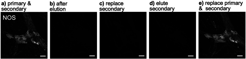
Indirect immunofluorescence is usually restricted to 3-5 markers per preparation, limiting analysis of coexistence. A solution containing 2-mercaptoethanol and sodium dodecyl sulfate (2-ME/SDS) can elute indirect immunofluorescence labelling (i.e. primary antisera followed by fluorophore-conjugated secondary antisera) and has been used for sequential staining of sections. The aim of this study was to test whether 2-ME/SDS is effective for eluting indirect immunofluorescent staining (with primary antisera visualised by fluorophore-coupled secondary antisera) in wholemount preparations. We also analysed how 2-ME/SDS may work and used this understanding to devise additional uses for immunofluorescence in the nervous system. 2-ME/SDS appears to denature unfixed proteins (including antisera used as reagents) but has much less effect on antigenicity of formaldehyde-fixed epitopes. Moieties linked by strong biotin-streptavidin bonds are highly resistant to elution by 2-ME/SDS. Two primary antisera raised in the same species can be applied without spurious cross-reactivity, if a specific order of labelling is followed. The first primary antiserum is followed by a biotinylated secondary, then a tertiary of fluorophore-conjugated streptavidin. The preparation is then exposed to 2-ME/SDS, which has minimal impact on labelling by the first primary/secondary/tertiary combination. However, when this is followed by a second primary antiserum (raised in the same species), followed by a fluorophore-conjugated secondary antiserum, the intervening 2-ME/SDS exposure prevents cross-reactivity between primary and secondary antisera of the two layers. A third property of 2-ME/SDS is that it reduces lipofuscin autofluorescence, although it also raises background fluorescence and strongly enhances autofluorescence of erythrocytes. In summary, 2-ME/SDS is easy to use, cost-effective and does not require modified primary antisera. It can be used as the basis of a multi-layer immunohistochemistry protocol and allows 2 primary antisera raised in the same species to be used together.
Keywords: Enteric nervous system; Fluorescent antibody technique; Indirect; Lipofuscin; Myenteric plexus; Protein denaturation.
© 2024 The Authors.
Conflict of interest statement
The authors declare that they have no known competing financial interests or personal relationships that could have appeared to influence the work reported in this paper.
Figures


Similar articles
-
Immunological reactivity of antisera prepared against the sodium dodecyl sulfate-treated structural polypeptides of adenovirus-associated virus.J Virol. 1972 Jun;9(6):1017-26. doi: 10.1128/JVI.9.6.1017-1026.1972. J Virol. 1972. PMID: 4338635 Free PMC article.
-
A simple method for immunofluorescent double staining with primary antisera from the same species.J Histochem Cytochem. 1993 Apr;41(4):627-30. doi: 10.1177/41.4.8450202. J Histochem Cytochem. 1993. PMID: 8450202
-
Elution of High-affinity (>10-9 KD) Antibodies from Tissue Sections: Clues to the Molecular Mechanism and Use in Sequential Immunostaining.J Histochem Cytochem. 2014 Jul;62(7):519-31. doi: 10.1369/0022155414536732. Epub 2014 May 2. J Histochem Cytochem. 2014. PMID: 24794148 Free PMC article.
-
Microwaving and Fluorophore-Tyramide for Multiplex Immunostaining on Mouse Adrenals - Using Unconjugated Primary Antibodies from the Same Host Species.J Vis Exp. 2020 Feb 21;(156):10.3791/60868. doi: 10.3791/60868. J Vis Exp. 2020. PMID: 32150172 Free PMC article.
-
Organization of the enteric nervous system in the human colon demonstrated by wholemount immunohistochemistry with special reference to the submucous plexus.Ann Anat. 1999 Jul;181(4):327-37. doi: 10.1016/S0940-9602(99)80122-8. Ann Anat. 1999. PMID: 10427369
References
-
- Zeng H., Sanes J.R. Neuronal cell-type classification: challenges, opportunities and the path forward. Nat. Rev. Neurosci. 2017;18(9):530–546. - PubMed
-
- Zeisel A., et al. Brain structure. Cell types in the mouse cortex and hippocampus revealed by single-cell RNA-seq. Science. 2015;347(6226):1138–1142. - PubMed
LinkOut - more resources
Full Text Sources
