

The relationship between epicardial adipose tissue volume on coronary computed tomography angiography and idiopathic ventricular tachycardia: a propensity score matching case-control study in Chinese population
- PMID: 38434552
- PMCID: PMC10904298
- DOI: 10.21037/cdt-23-345
The relationship between epicardial adipose tissue volume on coronary computed tomography angiography and idiopathic ventricular tachycardia: a propensity score matching case-control study in Chinese population
Abstract
Background: Large epicardial adipose tissue (EAT) volume is associated with the incidence of premature ventricular beats. The relationship between EAT volume and idiopathic ventricular tachycardia (IVT) is not yet clear. We aimed to investigate the effect of EAT volume on the risk of IVT.
Methods: This is a retrospective consecutive case-control study from January 2020 to September 2022. IVT patients (n=81) and control patients (n=162) undergoing coronary computed tomography angiography (CCTA) were retrospectively recruited. The patients in the control group were all hospitalized patients for different reasons, such as chest tightness, shortness of breath, chest pain, and so on. Demographic parameters and clinical characteristics of each individual were collected from the patient's medical records. We selected evaluation criteria for the conduct of a 1:1 propensity score (PS)-adjusted analysis. Multivariable logistic analysis was used to investigate risk factors for IVT. Furthermore, the impact of EAT volume on cardiac repolarization indices was assessed in IVT patients.
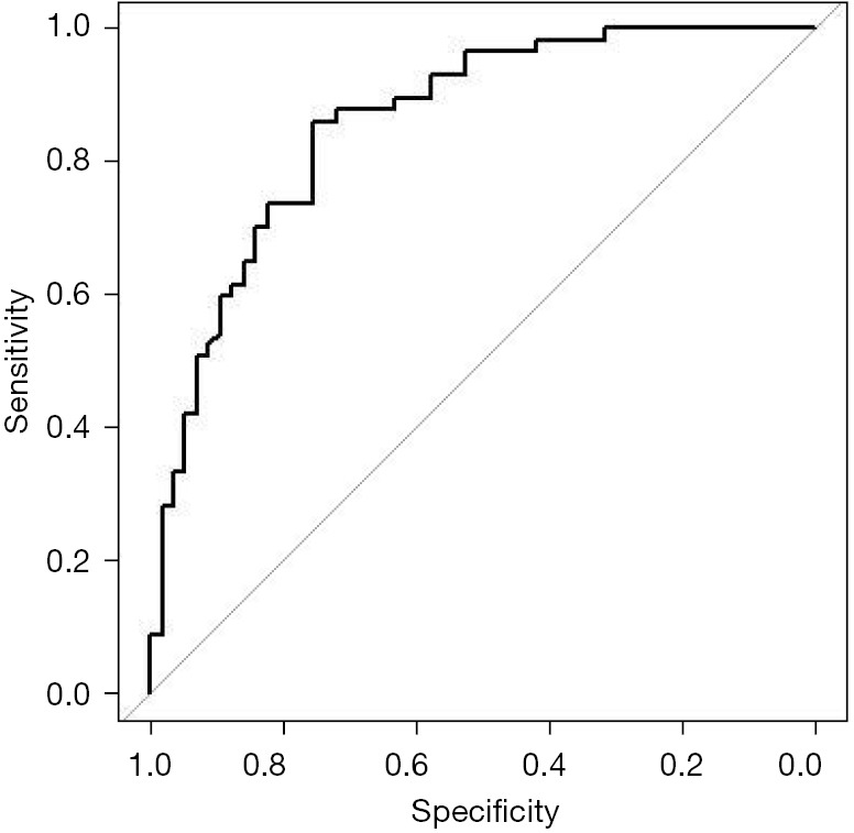
Results: Patients with IVT had a larger EAT volume than control group patients in the unadjusted cohort. Variables with P<0.10 in the univariable analysis and important factors were included in the multivariable analysis model, including body mass index (BMI), left ventricular ejection fraction (LVEF), early peak/artial peak (E/A) ratios <1, EAT attenuation, and EAT volume (per increase 10 mL). The multivariable logistic analysis found that EAT volume [per increase 10 mL, odds ratio (OR): 1.29, 95% confidence interval (CI): 1.17-1.41, P<0.001] was an independent risk factor for IVT. EAT volume (per increase 10 mL, OR: 1.43, 95% CI: 1.25-1.64, P<0.001) independent effect was demonstrated in the PS adjusted cohort (n=57 in both groups). The area under the curve of EAT volume to predict the risk of IVT patients in the PS adjusted cohort was 0.859. The sensitivity and specificity were 86.0%, and 75.4%, respectively. Furthermore, A large EAT volume of IVT patients had a longer time in Tp-e, and Tp-e/QTc, compared with low EAT volume.
Conclusions: Patients with IVT had increased EAT volume compared to control subjects. Our study revealed that large EAT volume is associated with an extended repolarization process in IVT patients. These insights are essential for understanding the mechanisms linking EAT with IVT.
Keywords: Idiopathic ventricular tachycardia (IVT); computed tomography (CT); electrocardiogram (ECG); epicardial adipose tissue (EAT).
2024 Cardiovascular Diagnosis and Therapy. All rights reserved.
Conflict of interest statement
Conflicts of Interest: All authors have completed the ICMJE uniform disclosure form (available at https://cdt.amegroups.com/article/view/10.21037/cdt-23-345/coif). The authors have no conflicts of interest to declare.
Figures


References
-
- Della Bella P, Baratto F, Vergara P, et al. Does Timing of Ventricular Tachycardia Ablation Affect Prognosis in Patients With an Implantable Cardioverter Defibrillator? Results From the Multicenter Randomized PARTITA Trial. Circulation 2022;145:1829-38. 10.1161/CIRCULATIONAHA.122.059598 - DOI - PubMed
LinkOut - more resources
Full Text Sources