. 2024 Feb 16;9(8):9735-9752.
doi: 10.1021/acsomega.3c09948. eCollection 2024 Feb 27.

Novel Chitosan-Coated Liposomes Coloaded with Exemestane and Genistein for an Effective Breast Cancer Therapy

Affiliations

Novel Chitosan-Coated Liposomes Coloaded with Exemestane and Genistein for an Effective Breast Cancer Therapy

Shwetakshi Sharma et al. ACS Omega. .

Abstract

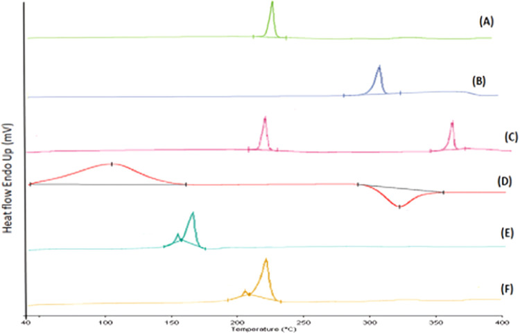
For achieving high effectiveness in the management of breast cancer, coadministration of drugs has attracted a lot of interest as a mode of therapy when compared to a single chemotherapeutic agent that often results in reduced therapeutic end results. Owing to their proven effectiveness, good patient compliance, and lower costs, oral anticancer drugs have received much attention. In the present work, we formulated the chitosan-coated nanoliposomes loaded with two lipophilic agents, namely, exemestane (EXE) and genistein (GEN). The formulation was prepared using the ethanol injection method, which is considered a simple method for getting the nanoliposomes. The formulation was optimized using Box-Behnken design (BBD) and was extensively characterized for particle size, ζ-potential, Fourier transform infrared (FTIR), differential scanning calorimetry (DSC), and X-ray diffraction (XRD) analysis. The sizes of conventional and coated liposomes were found to be 104.6 ± 3.8 and 120.3 ± 6.4 nm with a low polydispersity index of 0.399 and 0.381, respectively. The ζ-potential of the liposomes was observed to be -16.56 mV, which changed to a positive value of +22.4 mV, clearly indicating the complete coating of the nanoliposomes by the chitosan. The average encapsulation efficiency was found to be between 70 and 80% for all prepared formulations. The compatibility of the drug with excipients and complete dispersion of the drug inside the system were verified by FTIR, XRD, and DSC studies. Furthermore, the in vitro release studies concluded the sustained release pattern following the Korsmeyer-Peppas model as the best-fitting model with Fickian diffusion. Ex vivo studies showed better permeation of the chitosan-coated liposomes, which was further confirmed by confocal studies. The prepared chitosan-coated liposomes showed superior antioxidant activity (94.56%) and enhanced % cytotoxicity (IC50 7.253 ± 0.34 μM) compared to the uncoated liposomes. 3-(4,5-Dimethylthiazol-2-yl)-2,5-diphenyltetrazolium bromide (MTT) assay displayed better cytotoxicity of the chitosan-coated nanoliposomes compared to the plain drug, showing the better penetration and enhanced bioavailability of drugs inside the cells. The formulation was found to be safe for administration, which was confirmed using the toxicity studies performed on an animal model. The above data suggested that poorly soluble lipophilic drugs could be successfully delivered via chitosan-coated liposomes for their effective delivery in breast cancer.


Conflict of interest statement

The authors declare no competing financial interest.

Figures

Figure 1
Figure 1
Representation of three-dimensional plots displaying the effect of different independent variables on (A) particle size, (B) PDI, (C) % entrapment efficiency of EXE, and (D) % entrapment efficiency of GEN.
Figure 2
Figure 2
Particle size distribution of (A) placebo LIPO, (B) EXE-GEN-LIPO, (C) CS-EXE-GEN-LIPO, and ζ-potential of (D) placebo LIPO, (E) EXE-GEN-LIPO, and (F) CS-EXE-GEN-LIPO.
Figure 3
Figure 3
TEM micrographs of (A) optimized EXE-GEN-LIPO and (B) CS-EXE-GEN-LIPO (scale bar: 200 nm).
Figure 4
Figure 4
FTIR spectra of (A) EXE, (B) GEN, (C) physical mixture of drugs (EXE + GEN), (D) phospholipid, (E) chitosan, (F) optimized EXE-GEN-LIPO, and (G) optimized CS-EXE-GEN-LIPO.
Figure 5
Figure 5
DSC thermogram of (A) EXE, (B) GEN, (C) physical mixture of drugs (EXE + GEN), (D) chitosan, (E) lyophilized formulation of EXE-GEN-LIPO, and (F) lyophilized formulation of CS-EXE-GEN-LIPO.
Figure 6
Figure 6
XRD spectra of (A) EXE, (B) GEN, (C) cholesterol, (D) chitosan, (E) lyophilized formulation of EXE-GEN-LIPO, and (F) lyophilized formulation of CS-EXE-GEN-LIPO.
Figure 7
Figure 7
In vitro drug release profiles of EXE and GEN from EXE-SUSP, GEN-SUSP, EXE-GEN-SUSP, EXE-GEN-LIPO, and CS-EXE-GEN-LIPO at (A) pH 1.2 and (B) pH 6.8. The values are expressed as mean ± SD, n = 3.
Figure 8
Figure 8
Ex vivo intestinal permeation study demonstrating (A) cumulative amount of drug permeated (μg) versus time (min) and (B) cumulative amount of drug transported (μg/cm2) with time (min).
Figure 9
Figure 9
CLSM images of small intestine for determining the depth of penetration of (A) Rhodamine B, (B) Rhodamine B-labeled uncoated liposome, and (C) Rhodamine B-labeled coated liposome.
Figure 10
Figure 10
Histopathological photomicrographs of (A) heart, (B) liver, and (C) kidney of female Wistar rats treated with control, EXE-SUSP, GEN-SUSP, and CS-EXE-GEN-LIPO (40x).
Figure 11
Figure 11
Cellular uptake study performed on MCF-7 cells for plain Rhodamine B solution, Rhodamine B uncoated LIPO, and Rhodamine B Chitosan LIPO at 20×.
Figure 12
Figure 12
(A) Cell cytotoxicity (%) of MCF-7 cells treated with EXE-SUSP, GEN-SUSP, EXE-GEN-SUSP, EXE-GEN-LIPO, and CS-EXE-GEN-LIPO. (B) IC50 values obtained for EXE-SUSP, GEN-SUSP, EXE-GEN-SUSP, EXE-GEN-LIPO, and CS-EXE-GEN-LIPO. The results are expressed as percentage mean ± SD (n = 3) and *p < 0.05, **p < 0.01, ***p < 0.001 and ****p < 0.0001.
Figure 13
Figure 13
(A) Antioxidant activity of EXE-SUSP, GEN-SUSP, EXE-GEN-LIPO, CS-EXE-GEN-LIPO, and ascorbic acid as estimated by DPPH assay. (B) IC50 values obtained for EXE-SUSP, GEN-SUSP, EXE-GEN-LIPO, CS-EXE-GEN-LIPO, and ascorbic acid. The results are expressed as percentage mean ± SD (n = 3) and ***p < 0.001, ****p < 0.0001.
Figure 14
Figure 14
Particle size of (A) uncoated liposomes and (B) coated liposomes after 6 months of storage.

Similar articles

Cited by

References

    1. Arnold M.; Morgan E.; Rumgay H.; Mafra A.; Singh D.; Laversanne M.; Vignat J.; Gralow J. R.; Cardoso F.; Siesling S.; Soerjomataram I. Current and Future Burden of Breast Cancer: Global Statistics for 2020 and 2040. Breast 2022, 66, 15–23. 10.1016/j.breast.2022.08.010. - DOI - PMC - PubMed
    1. Sung H.; Ferlay J.; Siegel R. L.; Laversanne M.; Soerjomataram I.; Jemal A.; Bray F. Global Cancer Statistics 2020: GLOBOCAN Estimates of Incidence and Mortality Worldwide for 36 Cancers in 185 Countries. Ca-Cancer J. Clin. 2021, 71 (3), 209–249. 10.3322/caac.21660. - DOI - PubMed
    1. Anderson B. O.; Ilbawi A. M.; Fidarova E.; Weiderpass E.; Stevens L.; Abdel-Wahab M.; Mikkelsen B. The Global Breast Cancer Initiative: A Strategic Collaboration to Strengthen Health Care for Non-Communicable Diseases. Lancet Oncol. 2021, 22 (5), 578–581. 10.1016/S1470-2045(21)00071-1. - DOI - PubMed
    1. Kawish S. M.; Hasan N.; Beg S.; Qadir A.; Jain G. K.; Aqil M.; Ahmad F. J. Docetaxel-Loaded Borage Seed Oil Nanoemulsion with Improved Antitumor Activity for Solid Tumor Treatment: Formulation Development, in Vitro, in Silico and in Vivo Evaluation. J. Drug Delivery Sci. Technol. 2022, 75, 10369310.1016/j.jddst.2022.103693. - DOI
    1. Meng J.; Guo F.; Xu H.; Liang W.; Wang C.; Yang X.-D. Combination Therapy Using Co-Encapsulated Resveratrol and Paclitaxel in Liposomes for Drug Resistance Reversal in Breast Cancer Cells in Vivo. Sci. Rep. 2016, 6, 2239010.1038/srep22390. - DOI - PMC - PubMed

LinkOut - more resources