

doi: 10.1016/j.eats.2023.09.014.
eCollection 2024 Feb.
The Air-Lift Technique for Improving Ease and Safety When Establishing the Modified Midanterior Portal in Hip Arthroscopy
Affiliations
- PMID: 38435239
- PMCID: PMC10907895
- DOI: 10.1016/j.eats.2023.09.014
Item in Clipboard
The Air-Lift Technique for Improving Ease and Safety When Establishing the Modified Midanterior Portal in Hip Arthroscopy
Arthrosc Tech.
.
Abstract
Hip arthroscopy continues to increase in popularity and has an ever-expanding range of indications; however, the steep learning curve introduces significant risk of iatrogenic chondrolabral injury when accessing the joint and establishing arthroscopic portals. This article presents a technique for establishing the modified midanterior portal and is particularly useful when the available space is tight. We present "the air-lift" as a safe and simple adjunct to standard portal creation when performing hip arthroscopy in the supine position.
© 2023 The Authors.
Figures

Photograph of standard supine set up for hip arthroscopy using the Stryker Pivot Guardian hip distraction system and hip check (Stryker) with surface anatomy marked on the skin including the profile of the greater trochanter and a line extending distally from the anterior superior iliac spine. Patient's left side, supine position.

An air arthrogram is performed injecting air into the hip joint through a distal anterior location under image intensifier guidance (needle is labeled). After the joint has been insufflated, longitudinal traction is applied to the limb with approximately 75 lbs of force typically used to achieve distraction of the femoral head from the acetabulum. Patient's right side, supine position.

An anterior posterior view of the hip joint showing the initial needle for creation of the anterolateral viewing portal. Under radiograph guidance the initial anterolateral viewing portal is created with a needle followed by a wire positioned parallel to the sourcil of the acetabulum. Patient's right side, supine position.

An anterior posterior view of the hip joint showing the initial needle for creation of the anterolateral viewing portal. The initial needle is removed and the portal is dilated and developed. Following this a 70° arthroscope (Stryker, Denver, CO) is inserted. Patient's right side, supine position.
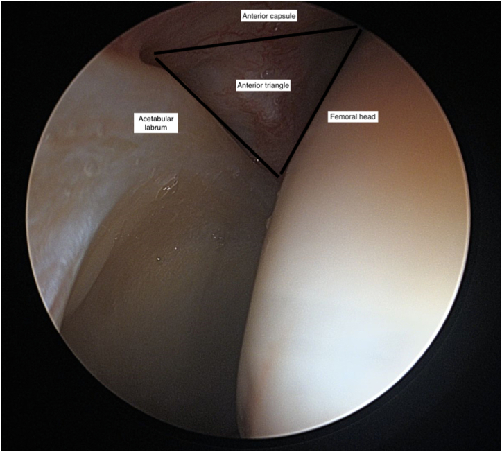
Surgeon’s view through the anterolateral portal with a 70° arthroscope visualizing the anterior triangle with the borders consisting of the acetabular labrum, femoral head and anterior capsule (labeled). This is performed as a dry view, as fluid has not yet been introduced to the hip joint. The desired entry point for the modified mid anterior portal is in the center of this triangle but this space can be tighter than ideal, with the needle and trocar risking chondral injury if we enter and then dilate in this location without sufficient space. Patient's left side, supine position.
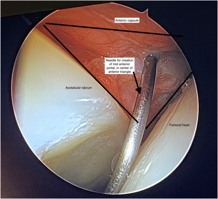
Surgeon’s view through the anterolateral viewing portal with the 70° arthroscope screen as the needle is inserted for creation of the mid anterior portal. The needle is in the center of the anterior triangle with the borders consisting of the acetabular labrum, femoral head and anterior capsule (labeled). Patient's left side, supine position.

Photograph of the setup for insufflation of the joint for the air-lift technique during supine hip arthroscopy. The arthroscope is in the anterolateral viewing portal with a 60-cc syringe attached to the 3-way mechanism on the arthroscope. This is then used to inflate the joint before creation of the modified midanterior portal. Please see related video for more information relating to this technique. Patient's right side, supine position.
References
-
- Ganz R., Parvizi J., Beck M., Leunig M., Notzli H., Siebenrock K.A. Femoroacetabular impingement: A cause for osteoarthritis of the hip. Clin Orthop Relat Res. 2003;417:112–120. - PubMed
-
- Maldonado D.R., Rosinsky P.J., Shapira J., Domb B.G. Stepwise safe access in hip arthroscopy in the supine position: Tips and pearls from A to Z. J Am Acad Orthop Surg. 2020;28:651–659. - PubMed
-
- Jimenez A.E., Monahan P.F., Owens J.S., Maldonado D.R., Curley A.J., Domb B.G. Earlier treatment yields superior outcomes in competitive athletes undergoing primary hip arthroscopy. Arthroscopy. 2021;38:2183–2191. - PubMed
-
- Maldonado D.R., Kufta A.Y., Krych A.J., et al. Primary hip arthroscopy for femoroacetabular impingement syndrome in adolescents improves outcomes and clinical benefit achievement rates at short term follow: A multicenter analysis. Arthroscopy. 2023;39:1211–1219. - PubMed
-
- Mehta N., Chamberlin P., Marx R.G., et al. Defining the learning curve for hip arthroscopy. Am J Sports Med. 2018;46:1285–1293. - PubMed
LinkOut - more resources
Full Text Sources
