Practical Aspects of Using Multifractal Formalism to Assess the Morphology of Biological Tissues
- PMID: 38435478
- PMCID: PMC10904356
- DOI: 10.17691/stm2023.15.3.04
Practical Aspects of Using Multifractal Formalism to Assess the Morphology of Biological Tissues
Abstract
The aim of the study is to identify practical aspects of using multifractal formalism to assess the morphology of biological tissues.
Materials and methods: The objects of the study were histological images of lung tissues of Wistar rats without pathology and with detected pathological changes, obtained at 50×, 100×, 200× magnifications. Image processing was carried out using the ImageJ/Fiji universal software. The multifractal spectrum of the images, processed to obtain a linear contour, was calculated with the use of FracLac - a module for ImageJ. This module was used to determine the scaling exponent (the function of the Rényi exponent, τ(q)) and the singularity spectrum itself.
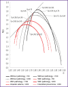
Results: The singularity spectra for tissues with no pathology have signs of multifractality. The image spectrum of tissue with pathology is shifted to the left relative to the spectrum characteristic of tissue without pathology. A decrease in the spectral height in the presence of pathology indicates a "simplification" of the alveolar pattern, which is presumably associated with the presence of widespread vasculitis, since it causes areas of hemorrhage to appear on the image; this leads to leveling the contour of the alveolar pattern, reducing the surface area of the alveoli and emerging areas inflamed by erythrocytes. At lower magnification, images with pathology lose signs of multifractality.
Conclusion: Correct results of assessing multifractal spectra of histological images can be achieved at 200× magnification and preprocessing to obtain linear contours. Significant differences between the morphological structure of lung tissues with and without pathology are observed when comparing the height, width, and position of the spectrum relative to the origin.
Keywords: alveolar pattern; fractal analysis; histology; image analysis; lung tissue.
Conflict of interest statement
Conflicts of interest. There are no conflicts of interest.
Figures


Similar articles
-
Multifractal analysis of uterine electromyography signals to differentiate term and preterm conditions.Proc Inst Mech Eng H. 2019 Mar;233(3):362-371. doi: 10.1177/0954411919827323. Epub 2019 Feb 1. Proc Inst Mech Eng H. 2019. PMID: 30706756
-
Classification of surface electromyographic signals by means of multifractal singularity spectrum.Med Biol Eng Comput. 2013 Mar;51(3):277-84. doi: 10.1007/s11517-012-0990-9. Epub 2012 Nov 7. Med Biol Eng Comput. 2013. PMID: 23132526
-
Impact of Healthy Aging on Multifractal Hemodynamic Fluctuations in the Human Prefrontal Cortex.Front Physiol. 2018 Aug 10;9:1072. doi: 10.3389/fphys.2018.01072. eCollection 2018. Front Physiol. 2018. PMID: 30147657 Free PMC article.
-
Multifractal detrended fluctuation analysis to characterize phase couplings in seahorse (Hippocampus kuda) feeding clicks.J Acoust Soc Am. 2014 Oct;136(4):1972-81. doi: 10.1121/1.4895713. J Acoust Soc Am. 2014. PMID: 25324096
-
Multifractal formalisms of human behavior.Hum Mov Sci. 2013 Aug;32(4):633-51. doi: 10.1016/j.humov.2013.01.008. Epub 2013 Aug 22. Hum Mov Sci. 2013. PMID: 24054900 Review.
References
-
- Huang H.K. Biomedical image processing. Crit Rev Bioeng. 1981;5(3):185–271. - PubMed
-
- Sokolova N.A., Orel V.E., Gusynin A.V., Selezneva A.A., Kolesnik S.V. Algorithm for image computerization analysis of histological specimens. Problemi informacijnih tehnologij. 2012;11:129–139.
MeSH terms
LinkOut - more resources
Full Text Sources
