

Anatomical Bases of the Temporal Muscle Trigger Points
- PMID: 38435541
- PMCID: PMC10908571
- DOI: 10.1155/2024/6641346
Anatomical Bases of the Temporal Muscle Trigger Points
Abstract
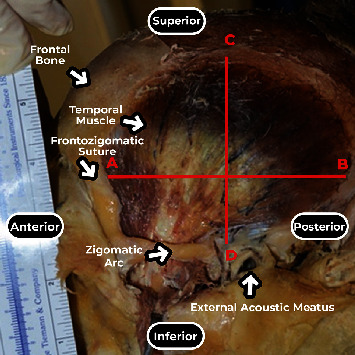
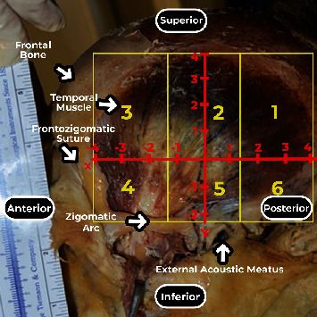
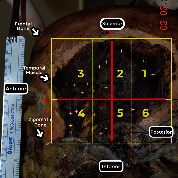
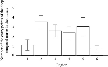
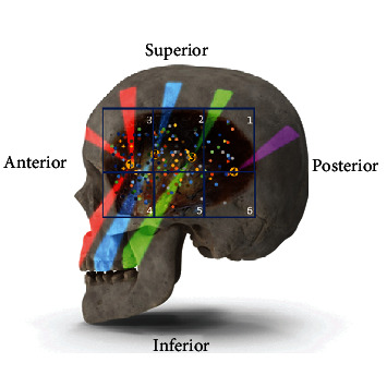
Method: Temporal muscles of 14 adult cadavers were studied. The muscle bellies were divided into six areas, three superior (1.2 and 3) and three inferior areas (4, 5, and 6) lower, according to a Cartesian plane to analyze and describe the entry points of the branches of the deep temporal nerves into the muscle. The branching distribution was analyzed using Poisson log-linear tests with Bonferroni post hoc tests for comparison between groups (sextants) (p < 0.05).
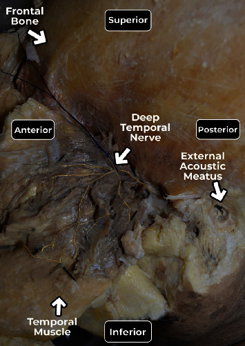
Results: Deep temporal nerve entry points were found in the temporal muscle in all areas. Most of the branches were observed in areas 2 and 5, which coincide with the muscle fibers responsible for mandible elevation and related to the previously described MTPs. Fewer branches were found in areas 1 and 6, where contraction produces mandible retraction.
Conclusion: There is an anatomical correlation between the branching pattern of the deep temporal nerve and temporal muscle trigger points. Adequate knowledge of the innervation of the temporal muscle may help elucidate the pathophysiology of myofascial syndromes and provide a rational basis for interventional or conservative approaches and help surgeons avoid iatrogenic lesions to the deep temporal nerve lesion.
Copyright © 2024 Luis Carlos Fernandez Garrido et al.
Conflict of interest statement
The authors have no financial or other potential conflicts of interest in the authorship or publication of this paper.
Figures

Similar articles
-
Anatomical Study of the Innervation of the Masseter Muscle and Its Correlation with Myofascial Trigger Points.J Pain Res. 2020 Dec 2;13:3217-3226. doi: 10.2147/JPR.S265717. eCollection 2020. J Pain Res. 2020. PMID: 33299345 Free PMC article.
-
An Anatomical Basis for the Myofascial Trigger Points of the Abductor Hallucis Muscle.Biomed Res Int. 2020 Jan 22;2020:9240581. doi: 10.1155/2020/9240581. eCollection 2020. Biomed Res Int. 2020. PMID: 32076620 Free PMC article.
-
Trigger points: an anatomical substratum.Biomed Res Int. 2015;2015:623287. doi: 10.1155/2015/623287. Epub 2015 Feb 24. Biomed Res Int. 2015. PMID: 25811029 Free PMC article.
-
Myofascial trigger point pain.Alpha Omegan. 2013 Spring-Summer;106(1-2):14-22. Alpha Omegan. 2013. PMID: 24864393 Review.
-
Latent myofascial trigger points.Curr Pain Headache Rep. 2011 Oct;15(5):386-92. doi: 10.1007/s11916-011-0210-6. Curr Pain Headache Rep. 2011. PMID: 21559783 Review.
References
-
- De la Torre Canales G., Poluha R. L., Pinzón N. A., et al. Efficacy of botulinum toxin type-A I in the improvement of mandibular motion and muscle sensibility in myofascial pain TMD subjects: a randomized controlled trial. Toxins . 2022;14(7):p. 441. doi: 10.3390/toxins14070441. - DOI - PMC - PubMed
MeSH terms
LinkOut - more resources
Full Text Sources
