

Machine learning based prediction of image quality in prostate MRI using rapid localizer images
- PMID: 38435711
- PMCID: PMC10905647
- DOI: 10.1117/1.JMI.11.2.026001
Machine learning based prediction of image quality in prostate MRI using rapid localizer images
Abstract
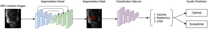
Purpose: Diagnostic performance of prostate MRI depends on high-quality imaging. Prostate MRI quality is inversely proportional to the amount of rectal gas and distention. Early detection of poor-quality MRI may enable intervention to remove gas or exam rescheduling, saving time. We developed a machine learning based quality prediction of yet-to-be acquired MRI images solely based on MRI rapid localizer sequence, which can be acquired in a few seconds.
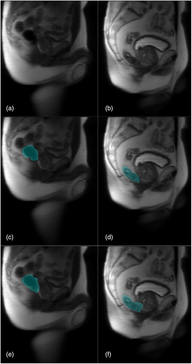
Approach: The dataset consists of 213 (147 for training and 64 for testing) prostate sagittal T2-weighted (T2W) MRI localizer images and rectal content, manually labeled by an expert radiologist. Each MRI localizer contains seven two-dimensional (2D) slices of the patient, accompanied by manual segmentations of rectum for each slice. Cascaded and end-to-end deep learning models were used to predict the quality of yet-to-be T2W, DWI, and apparent diffusion coefficient (ADC) MRI images. Predictions were compared to quality scores determined by the experts using area under the receiver operator characteristic curve and intra-class correlation coefficient.
Results: In the test set of 64 patients, optimal versus suboptimal exams occurred in 95.3% (61/64) versus 4.7% (3/64) for T2W, 90.6% (58/64) versus 9.4% (6/64) for DWI, and 89.1% (57/64) versus 10.9% (7/64) for ADC. The best performing segmentation model was 2D U-Net with ResNet-34 encoder and ImageNet weights. The best performing classifier was the radiomics based classifier.
Conclusions: A radiomics based classifier applied to localizer images achieves accurate diagnosis of subsequent image quality for T2W, DWI, and ADC prostate MRI sequences.
Keywords: artifact; gas; magnetic resonance imaging MRI; prostate; quality.
© 2024 Society of Photo-Optical Instrumentation Engineers (SPIE).
Figures
Similar articles
-
Fully automated detection of prostate transition zone tumors on T2-weighted and apparent diffusion coefficient (ADC) map MR images using U-Net ensemble.Med Phys. 2021 Nov;48(11):6889-6900. doi: 10.1002/mp.15181. Epub 2021 Aug 30. Med Phys. 2021. PMID: 34418108
-
Segmentation of the prostate, its zones, anterior fibromuscular stroma, and urethra on the MRIs and multimodality image fusion using U-Net model.Quant Imaging Med Surg. 2022 Oct;12(10):4786-4804. doi: 10.21037/qims-22-115. Quant Imaging Med Surg. 2022. PMID: 36185056 Free PMC article.
-
The Effect of Image Resampling on the Performance of Radiomics-Based Artificial Intelligence in Multicenter Prostate MRI.J Magn Reson Imaging. 2024 May;59(5):1800-1806. doi: 10.1002/jmri.28935. Epub 2023 Aug 12. J Magn Reson Imaging. 2024. PMID: 37572098
-
Automated segmentation of prostate zonal anatomy on T2-weighted (T2W) and apparent diffusion coefficient (ADC) map MR images using U-Nets.Med Phys. 2019 Jul;46(7):3078-3090. doi: 10.1002/mp.13550. Epub 2019 May 11. Med Phys. 2019. PMID: 31002381
-
3.0 T prostate MRI: Visual assessment of 2D and 3D T2-weighted imaging sequences using PI-QUAL score.Eur J Radiol. 2023 Sep;166:110974. doi: 10.1016/j.ejrad.2023.110974. Epub 2023 Jul 12. Eur J Radiol. 2023. PMID: 37453273
Cited by
-
Insights into Radiology Publications.Indian J Radiol Imaging. 2025 Jan 9;35(Suppl 1):S18-S29. doi: 10.1055/s-0044-1793914. eCollection 2025 Jan. Indian J Radiol Imaging. 2025. PMID: 39802728 Free PMC article.
-
Optimizing the early diagnosis of neurological disorders through the application of machine learning for predictive analytics in medical imaging.Sci Rep. 2025 Jul 2;15(1):22488. doi: 10.1038/s41598-025-05888-z. Sci Rep. 2025. PMID: 40594215 Free PMC article.
References
LinkOut - more resources
Full Text Sources
