. 2023 Aug 2;9(2):e10584.
doi: 10.1002/btm2.10584. eCollection 2024 Mar.

A drug-selectable acoustic reporter gene system for human cell ultrasound imaging

Affiliations

A drug-selectable acoustic reporter gene system for human cell ultrasound imaging

Alessandro R Howells et al. Bioeng Transl Med. .

Abstract

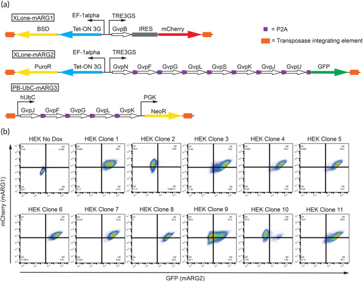
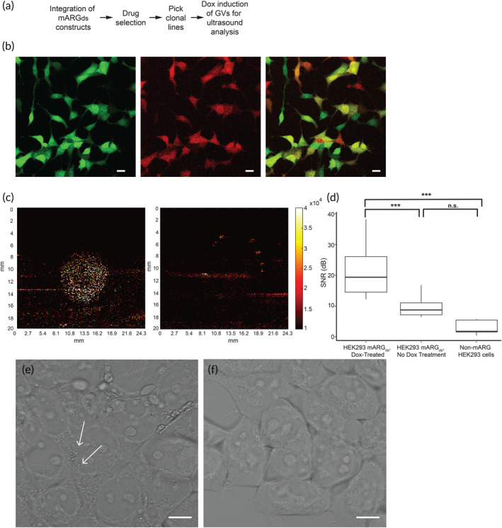
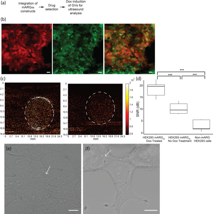
A promising new field of genetically encoded ultrasound contrast agents in the form of gas vesicles has recently emerged, which could extend the specificity of medical ultrasound imaging. However, given the delicate genetic nature of how these genes are integrated and expressed, current methods of producing gas vesicle-expressing mammalian cell lines requires significant cell processing time to establish a clonal/polyclonal line that robustly expresses the gas vesicles sufficiently enough for ultrasound contrast. Here, we describe an inducible and drug-selectable acoustic reporter gene system that can enable gas vesicle expression in mammalian cell lines, which we demonstrate using HEK293T cells. Our drug-selectable construct design increases the stability and proportion of cells that successfully integrate all plasmids into their genome, thus reducing the amount of cell processing time required. Additionally, we demonstrate that our drug-selectable strategy forgoes the need for single-cell cloning and fluorescence-activated cell sorting, and that a drug-selected mixed population is sufficient to generate robust ultrasound contrast. Successful gas vesicle expression was optically and ultrasonically verified, with cells expressing gas vesicles exhibiting an 80% greater signal-to-noise ratio compared to negative controls and a 500% greater signal-to-noise ratio compared to wild-type HEK293T cells. This technology presents a new reporter gene paradigm by which ultrasound can be harnessed to visualize specific cell types for applications including cellular reporting and cell therapies.

Keywords: acoustic reporter genes; cell engineering; ultrasound imaging.


Conflict of interest statement

The authors declare no conflict of interest.

Figures

FIGURE 1
FIGURE 1
Design of mARGds HEK293T clones. (a) Schematic of the transposase integrating mARGds construct designs. From left to right, the XLone‐mARG1 cassette possesses the Blasticidin resistant gene and the Tet‐On 3G system downstream of the constitutive EF‐1α promoter, and the mARG GvpB gene and mCherry downstream of the Dox inducible TRE3GS promoter. The XLone‐mARG2 cassette possesses the Puromycin resistant gene and the Tet‐On 3G system downstream of the constitutive EF‐1α promoter, and the mARG GvpF, GvpG, GvpL, GvpS, GvpK, GvpJ, GvpU genes (separated by P2A) and GFP downstream of the Dox inducible TRE3GS promoter. Finally, PB‐UbC‐mARG3 cassette possesses the mARG GvpF, GvpG, GvpL, and GvpK genes downstream of the constitutive hUbC promoter and Geneticin resistant gene downstream of the constitutive PGK promoter. (b) Flow cytometry of mARGds HEK293T clonal lines against GFP (XLone‐mARG2) and mCherry (XLone‐mARG1).
FIGURE 2
FIGURE 2
Single cell mARGds HEK293T cells express gas vesicles and produce ultrasound contrast. (a) Process flow for generating gas vesicle‐producing single‐cell clone HEK293T cell lines. (b) Clone 6 HEK293T mARGds gene expression validated by fluorescence microscopy. Scale bar = 10 μm. (c) Ultrasound contrast is produced by doxycycline‐treated HEK293T mARGds single cell clones after 3 days of treatment (left panel) whereas cells not treated with doxycycline produce no ultrasound contrast (right panel). (d) Signal to noise ratio of ultrasound imaging is significantly stronger in doxycycline‐treated cells compared to non‐doxycycline treated cells and non‐mARGds cells, indicative of gas vesicle production (n = 6). (e) Phase contrast image of HEK293T mARGds cells after 3 days of doxycycline treatment with small puncta spread throughout the cell (white arrows), indicative of gas vesicle presence. (f) Phase contrast image of HEK293T mARGds cells undergoing no doxycycline treatment.
FIGURE 3
FIGURE 3
Mixed population HEK293T mARGds cells produce sufficient ultrasound contrast after several days of antibiotic selection and doxycycline treatment. (a) Process flow for generating gas vesicle‐producing mixed population HEK293T cells. (b) Antibiotic selected and doxycycline‐treated HEK293T mARGds mixed population cells properly integrated the mARGds genes. Scale bar = 10 μm. (c) Doxycycline‐treated HEK293T mARGds mixed population cells produce stronger ultrasound contrast (left panel) compared to non‐doxycycline treated mARGds mixed population cells. (d) mARGds mixed population HEK293T cells exhibit a stronger signal to noise (SNR) ratio after 3 days of doxycycline treatment compared to non‐doxycycline treated mARGds cells and non‐mARG cells (n = 7). (e) Phase contrast image of HEK293T mARGds cells after 24 h of doxycycline treatment with small puncta spread throughout the cell (white arrows), indicative of gas vesicle presence. (f) Phase contrast image of HEK293T mARGds cells undergoing no doxycycline treatment. Small puncta are still present (white arrows), though in much smaller quantities than doxycycline‐treated cells.

References

    1. Chalfie M, Tu Y, Euskirchen G, Ward WW, Prasher DC. Green fluorescent protein as a marker for gene expression. Science. 1994;263(5148):802‐805. - PubMed
    1. Fernández‐Suárez M, Ting AY. Fluorescent probes for super‐resolution imaging in living cells. Nat Rev Mol Cell Biol. 2008;9(12):929‐943. - PubMed
    1. Chalfie M. Green fluorescent protein. Photochem Photobiol. 1995;62(4):651‐656. - PubMed
    1. Feng G, Mellor RH, Bernstein M, et al. Imaging neuronal subsets in transgenic mice expressing multiple spectral variants of GFP. Neuron. 2000;28(1):41‐51. - PubMed
    1. Jensen EC. Use of fluorescent probes: their effect on cell biology and limitations. Anat Rec. 2012;295(12):2031‐2036. - PubMed

LinkOut - more resources