Review
. 2024 Jan 11;9(2):e10623.
doi: 10.1002/btm2.10623. eCollection 2024 Mar.

Micro Trojan horses: Engineering extracellular vesicles crossing biological barriers for drug delivery

Affiliations
Review

Micro Trojan horses: Engineering extracellular vesicles crossing biological barriers for drug delivery

Bin Zeng et al. Bioeng Transl Med. .

Abstract

The biological barriers of the body, such as the blood-brain, placental, intestinal, skin, and air-blood, protect against invading viruses and bacteria while providing necessary physical support. However, these barriers also hinder the delivery of drugs to target tissues, reducing their therapeutic efficacy. Extracellular vesicles (EVs), nanostructures with a diameter ranging from 30 nm to 10 μm secreted by cells, offer a potential solution to this challenge. These natural vesicles can effectively pass through various biological barriers, facilitating intercellular communication. As a result, artificially engineered EVs that mimic or are superior to the natural ones have emerged as a promising drug delivery vehicle, capable of delivering drugs to almost any body part to treat various diseases. This review first provides an overview of the formation and cross-species uptake of natural EVs from different organisms, including animals, plants, and bacteria. Later, it explores the current clinical applications, perspectives, and challenges associated with using engineered EVs as a drug delivery platform. Finally, it aims to inspire further research to help bioengineered EVs effectively cross biological barriers to treat diseases.

Keywords: biological barriers; drug delivery; engineering; extracellular vesicles; therapy.


Conflict of interest statement

The authors have no conflicts of interest to declare.

Figures

FIGURE 1
FIGURE 1
Sketch of the generation and cross‐species uptake of extracellular vesicles. The figure demonstrates different mechanisms involved in generating and releasing vesicles in animal cells, plant cells, and bacteria. Animal cell‐derived vesicles are mainly caused by forming multi‐vesicular bodies (MVBs) and released through ESCRT‐dependent or ESCRT‐independent pathways. Alternatively, vesicles can be produced by plasma membrane budding or IPMC‐mediated release. Plant cells also produce and uptake vesicles, but they use a unique organelle called EXPO to produce vesicles. Moreover, vesicles produced by plant cells need to cross the cell wall, and the process and mechanism of vesicles crossing the plant cell wall remain unclear. Bacterial vesicles are produced through membrane budding and bacterial autolysis. The mechanism of production and contents of Gram‐negative bacterial vesicles (lower right) are associated with lipid and protein accumulation in the periplasm and outer lobules, flagellar assembly, and bacterial autolysis due to stress and infection. In contrast, positive bacterial vesicles (lower left) are produced through plasma membrane lipid accumulation and bacterial autolysis. The middle part of the figure displays the signature proteins and contents of vesicles from animals, plants, and bacteria.
FIGURE 2
FIGURE 2
Illustration of the methods for isolating extracellular vesicles. (a) These methods can be categorized into three groups based on their principles: separation methods using differential centrifugation (e.g., ultracentrifugation and density gradient centrifugation), separation methods based on size (e.g., ultrafiltration, tangential flow filtration), and methods based on antigen–antibody specificity (e.g., immunoaffinity capture). (b) Representative data of extracellular vesicle identification include nanoparticle size determination with nanoparticle tracking analysis (NTA), morphology identification with transmission electron microscopy (TEM), and western blot identification of specific marker protein expression.
FIGURE 3
FIGURE 3
Illustration of the various techniques used for engineering vesicle modifications. (a) These engineering techniques for extracellular vesicles can be classified into physical, biological, and chemical modifications. Physical modification techniques such as extrusion, ultrasonication, and electroporation can be employed for membrane fusion, drug loading, and other applications. Biomodification involves genetically engineering cells to express specific products, which are then carried by the vesicles produced by these cells. Chemical modification methods such as covalent coupling, lipophilic insertion, and receptor‐ligand binding are also used to modify extracellular vesicles. (b) Click chemistry, which involves the covalent bonding of azides and alkynes, is commonly used in nanoparticle engineering. (c) Genetic engineering techniques can be used to load extracellular vesicles with genetically encoded nucleic acids, proteins, polypeptides, and glycocalyx in their surface or cavity. Commonly used extracellular vesicle proteins for genetic engineering include CD63, CD9, Lamp2b, and ClyA in bacterial vesicles. (d) After isolation and purification, natural or genetically engineered extracellular vesicles can be modified using physical or chemical methods to enhance their targeting and drug‐loading capabilities. Several clinical trials based on engineered extracellular vesicles are currently underway, focusing on treating diseases such as COVID‐19, cancer, and others.
FIGURE 4
FIGURE 4
Schematic diagram of the mechanism of bacterial vesicles crossing the intestinal barrier. As shown in the upper part of the figure, vesicles from multiple origins could cross the intestinal barrier with good potential for drug delivery. The middle part of the figure summarizes the mechanism by which extracellular vesicles cross the intestinal endothelial barrier. The mechanisms for crossing the barrier are mainly divided into three categories: (1) transcytosis; (2) intercellular pathway; (3) DC cell extension pseudopodia. The lower part of the figure summarizes the regulation of immune cells by extracellular vesicles from different sources. Among them, the regulatory function of BEV on immune cells is bidirectional. Some BEVs can promote the differentiation of pro‐inflammatory immune cells (e.g., Th1, Th17), while others can promote the differentiation of anti‐inflammatory immune cells (e.g., Th2, Treg cells). MSCs and plant‐derived EVs mainly inhibit the secretion of various inflammatory cytokines and promote the anti‐inflammatory phenotype differentiation of immune cells.
FIGURE 5
FIGURE 5
Schematic diagram of gut microbiota vesicles crossing the intestinal barrier to regulate other organ functions. (a) Extracellular vesicles originating from the intestinal microbiota can traverse the intestinal barrier and access other organs within the human body, thus playing a role in the pathogenesis of various diseases. Specific bacterial extracellular vesicles have a protective effect on the intestine and other organs. In contrast, others can compromise intestinal homeostasis, disrupt endothelial barrier integrity, and incite inflammatory responses resulting in organ damage. These pro‐inflammatory bacterial vesicles may be useful in disease diagnosis or represent attractive therapeutic targets. (b) Once extracellular vesicles (EVs) have crossed the intestinal barrier, extracellular vesicles can penetrate the blood–brain barrier and enter the central nervous system, potentially serving a regulatory function through transcytosis and paracellular pathways. (c) Children's gut microbiota‐derived extracellular vesicles (CGM‐EVs) of the gut microbiota in children are believed to facilitate bone formation while inhibiting bone resorption, playing a crucial role in maintaining the balance of bone metabolism.
FIGURE 6
FIGURE 6
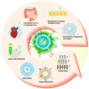
Conclusion and perspectives. Extracellular vesicles (EVs) have emerged as a promising drug delivery system (DDS) for targeted therapy by crossing biological barriers. Engineering modifications can enhance EVs' penetration ability, but toxicity and immune system activation must be avoided. Optimizing EV separation and purification can increase their homogeneity and clinical potential. In addition, further research on EV subpopulations and production pathways can improve their characterization and identification. Taken together, EVs show significant potential as a DDS but require continued optimization for clinical application.

Similar articles

Cited by

References

    1. Weibel ER, Knight BW. A morphometric study on the thickness of the pulmonary air‐blood barrier. J Cell Biol. 1964;21:367‐384. - PMC - PubMed
    1. Harris‐Tryon TA, Grice EA. Microbiota and maintenance of skin barrier function. Science. 2022;376:940‐945. - PubMed
    1. Arumugasaamy N, Rock KD, Kuo C‐Y, Bale TL, Fisher JP. Microphysiological systems of the placental barrier. Adv Drug Deliv Rev. 2020;161–162:161‐175. - PMC - PubMed
    1. Camilleri M. Leaky gut: mechanisms, measurement and clinical implications in humans. Gut. 2019;68:1516‐1526. - PMC - PubMed
    1. Kreyling WG, Hirn S, Möller W, et al. Air–blood barrier translocation of tracheally instilled gold nanoparticles inversely depends on particle size. ACS Nano. 2014;8:222‐233. - PMC - PubMed