

Placental Transmogrification of the Lung in a Patient Without Emphysematous Disease
- PMID: 38435917
- PMCID: PMC10906123
- DOI: 10.7759/cureus.53294
Placental Transmogrification of the Lung in a Patient Without Emphysematous Disease
Abstract
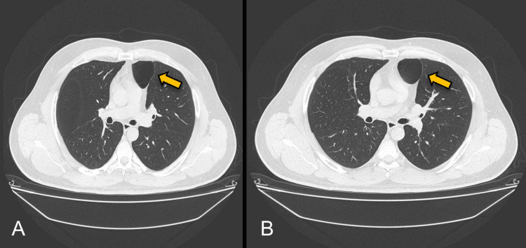
We present a 47-year-old male without a relevant history or past respiratory diseases. He debuted with an acute, non-complicated COVID-19 infection, and later he started with mMRC-2 dyspnea, accompanied by a non-expectorant cough of four months evolution. A CT thoracic scan showed a dilatation of the aerial homogenous space and a well-defined anterior left pericardiac level, and a pericardial left bulla was diagnosed. The patient was treated with surgical intervention by video-assisted thoracoscopic surgery and had an adequate post-surgical evolution. The PPT must be managed by a multidisciplinary team with the definitive treatment of surgical resection.
Keywords: minimally invasive lung resection; pulmonary bulla; pulmonary placental transmogrification; thoracic ct scan; thorax surgery.
Copyright © 2024, Arias Rivera et al.
Conflict of interest statement
The authors have declared that no competing interests exist.
Figures


References
-
- Placental transmogrification of the lung. Kim JW, Park IH, Kwon W, Eom MS, Kim YJ, Oh JH. https://doi.org/10.3348/kjr.2013.14.6.977. Korean J Radiol. 2013;14:977–980. - PMC - PubMed
-
- Pulmonary placental transmogrification: the last 16 years in a reference centre. Ortiz S, Tortosa F. https://doi.org/10.1016/j.rppnen.2017.02.007. Rev Port Pneumol (2006) 2017;23:164–166. - PubMed
-
- Unilateral giant bullous emphysema with placental transmogrification of the lung. Horsley WS, Gal AA, Mansour KA. Ann Thorac Surg. 1997;64:226–228. - PubMed
-
- [Placental transmogrification of the lung. Atypical presentation of the bullous emphysema] Vila L, Reginatto A, Rivero H, Rayá M, Guma G, Patané AK. https://pubmed.ncbi.nlm.nih.gov/33048806/ Medicina (B Aires) 2020;80:570–573. - PubMed
-
- Placental transmogrification of the lung. Foschini G, Rodríguez CM, Rubio MM, Baldo X. https://doi.org/10.1016/j.arbres.2021.04.004. Arch Bronconeumol. 2022;58:433. - PubMed
Publication types
LinkOut - more resources
Full Text Sources
Miscellaneous