. 2024 Apr 16;98(4):e0184423.
doi: 10.1128/jvi.01844-23. Epub 2024 Mar 4.

GTPase activity of porcine Mx1 plays a dominant role in inhibiting the N-Nsp9 interaction and thus inhibiting PRRSV replication

Affiliations

GTPase activity of porcine Mx1 plays a dominant role in inhibiting the N-Nsp9 interaction and thus inhibiting PRRSV replication

Yue Hu et al. J Virol. .

Abstract

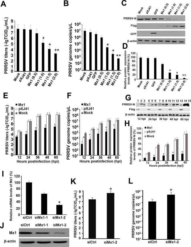
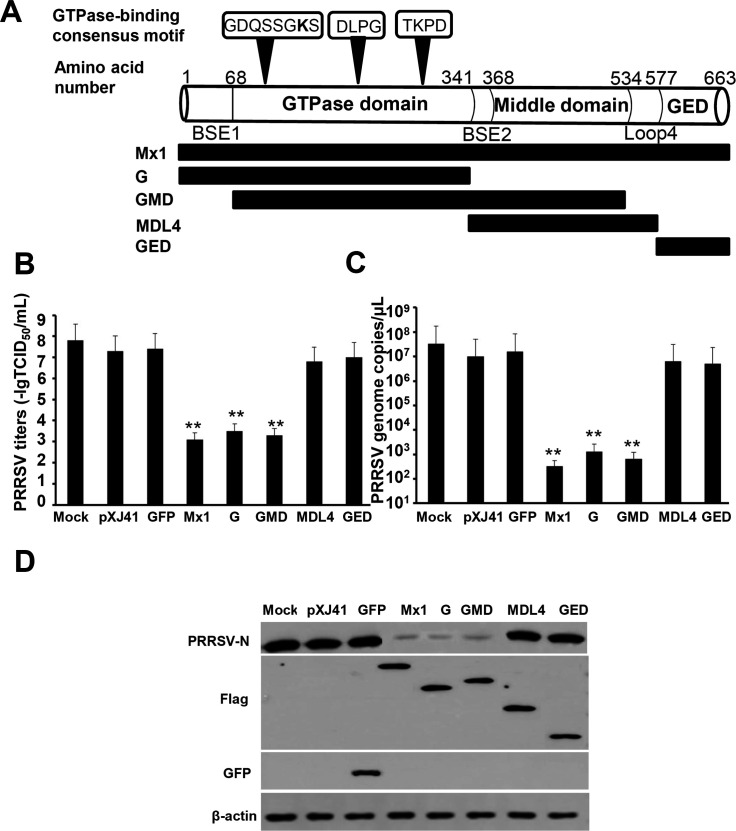
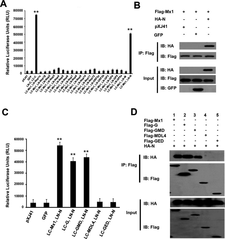
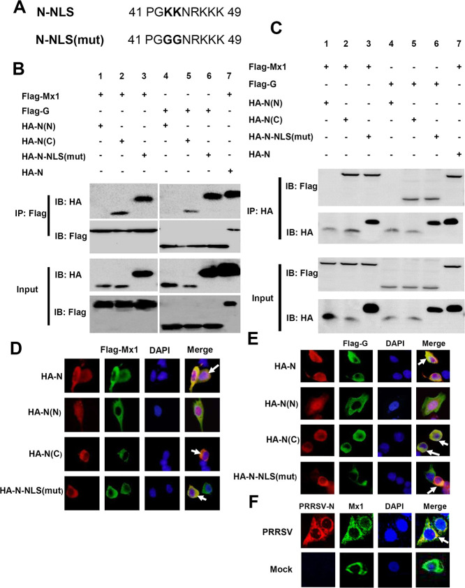
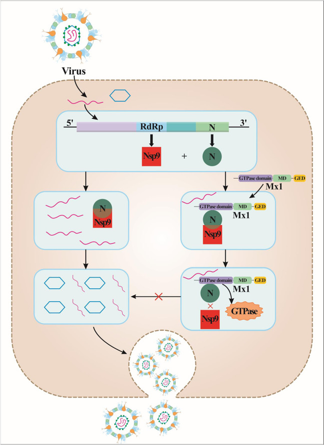
Porcine Mx1 is a type of interferon-induced GTPase that inhibits the replication of certain RNA viruses. However, the antiviral effects and the underlying mechanism of porcine Mx1 for porcine reproductive and respiratory syndrome virus (PRRSV) remain unknown. In this study, we demonstrated that porcine Mx1 could significantly inhibit PRRSV replication in MARC-145 cells. By Mx1 segment analysis, it was indicated that the GTPase domain (68-341aa) was the functional area to inhibit PRRSV replication and that Mx1 interacted with the PRRSV-N protein through the GTPase domain (68-341aa) in the cytoplasm. Amino acid residues K295 and K299 in the G domain of Mx1 were the key sites for Mx1-N interaction while mutant proteins Mx1(K295A) and Mx1(K299A) still partially inhibited PRRSV replication. Furthermore, we found that the GTPase activity of Mx1 was dominant for Mx1 to inhibit PRRSV replication but was not essential for Mx1-N interaction. Finally, mechanistic studies demonstrated that the GTPase activity of Mx1 played a dominant role in inhibiting the N-Nsp9 interaction and that the interaction between Mx1 and N partially inhibited the N-Nsp9 interaction. We propose that the complete anti-PRRSV mechanism of porcine Mx1 contains a two-step process: Mx1 binds to the PRRSV-N protein and subsequently disrupts the N-Nsp9 interaction by a process requiring the GTPase activity of Mx1. Taken together, the results of our experiments describe for the first time a novel mechanism by which porcine Mx1 evolves to inhibit PRRSV replication.

Importance: Mx1 protein is a key mediator of the interferon-induced antiviral response against a wide range of viruses. How porcine Mx1 affects the replication of porcine reproductive and respiratory syndrome virus (PRRSV) and its biological function has not been studied. Here, we show that Mx1 protein inhibits PRRSV replication by interfering with N-Nsp9 interaction. Furthermore, the GTPase activity of porcine Mx1 plays a dominant role and the Mx1-N interaction plays an assistant role in this interference process. This study uncovers a novel mechanism evolved by porcine Mx1 to exert anti-PRRSV activities.

Keywords: Mx1; antiviral agents; interferons; porcine reproductive and respiratory syndrome virus.


Conflict of interest statement

The authors declare no conflict of interest.

Figures

Fig 1
Fig 1
Mx1 protein could inhibit PRRSV replication in MARC-145 and PAM-KNU cells. (A–C) MARC-145 cells were seeded in 6-well plates and grown to 60% to 70% confluence. Flag-Mx1 (from 0.5 to 2.0 µg), pXJ41 empty vector (1.0 µg), GFP plasmid (1.0 µg), or mock was transfected into the cells. At 24-h posttransfection, the cells were infected with HP-PRRSV SY0608 strain at an MOI of 0.1. At 48-h postinfection, the supernatants of the cells were harvested and the whole-cell lysates were collected. TCID50 of PRRSV (A) and PRRSV genome copies (B) were detected, respectively, as described in the Materials and Methods. (C) PRRSV-N, Mx1, GFP, and β-actin expression were detected in the lysates by Western blotting as described in the Materials and Methods. The data presented here are results from one experiment of three Western blotting experiments. (D) Densitometry analysis of the digital image of PRRSV-N in Western blotting from three independent experiments. The band intensities of blotting with PRRSV-N specific monoclonal antibody SDOW-17 are shown as the relative protein expression levels, normalized with β-actin. (E–G) MARC-145 cells were seeded in 6-well plates and grown to 60% to 70% confluence. Flag-Mx1 (1.0 µg), pXJ41 empty vector (1.0 µg), or mock was transfected into the cells. At 24-h posttransfection, the cells were infected with HP-PRRSV SY0608 strain at an MOI of 0.1. At indicated postinfection time (from 12 to 60 h), the supernatants of the cells were harvested and the whole-cell lysates were collected. TCID50 of PRRSV (E) and PRRSV genome copies (F) were detected, respectively, as described in the Materials and Methods. (G) The expression of PRRSV-N in Mx1-transfected cells (lanes 1, 4, 7, 10, 13), pXJ41-transfected cells (lanes 2, 5, 8, 11, 14) , and mock-transfected cells (lanes 3, 6, 9, 12, 15), and β-actin expression were detected in different groups of cell lysates by Western blotting as described in the Materials and Methods. The data presented here are results from one experiment of three Western blotting experiments. (H) Densitometry analysis of the digital image of PRRSV-N in Western blotting from three independent experiments. The band intensities of blotting with PRRSV-N specific monoclonal antibody SDOW-17 are shown as the relative protein expression levels, normalized with β-actin. (I) Real-time RT-PCR analysis of the efficiency of siRNA targeting Mx1 in PAM-KNU cells. Cells were transfected with siRNA (siMx1-1, siMx1-2) or negative control siRNA (siCtrl). The total cellular RNA was extracted at 24-h posttransfection and analyzed by real-time RT-PCR. Relative transcript levels of Mx1 are shown as relative percentage changes in comparison with the level for siCtrl-transfected cells. (J) Western blotting analysis of siRNA-mediated Mx1 knockdown cells. Lysates of PAM-KNU cells transfected with siRNA (siMx1-1, siMx1-2) or siCtrl were prepared at 24-h posttransfection and subjected to Western blotting. Endogenous Mx1 was detected using anti-Mx1 antibody. β-actin was used as a loading control. (K) TCID50 of PRRSV in siMx1-2- or siCtrl-transfected PAM-KNU cells. Cells were transfected with siMx1-2 or siCtrl for 24 h, followed by infection with HP-PRRSV SY0608 strain at an MOI of 0.1 for 36 h; TCID50 was detected as described in the Materials and Methods. (L) Absolute quantitative RT-PCR analysis of PRRSV genome copies in siMx1-2- or siCtrl-transfected PAM-KNU cells. PRRSV genome copies were also detected as described above. Data represent the means ± standard deviations (error bars) of three independent experiments. *, P < 0.05; **, P < 0.01.
Fig 2
Fig 2
GTPase domain (68-341aa) was the functional area for Mx1 to inhibit PRRSV replication. (A) The schematic representation of Mx1 protein domains and their positions with indications of various truncations of Mx1 protein used in this study. (B–C) MARC-145 cells were transfected with Flag-G, Flag-GMD, Flag-MDL4, Flag-GED, pXJ41 empty vector, GFP plasmid, or mock-transfected. TCID50 of PRRSV (B) and PRRSV genome copies (C) were detected as described in the Materials and Methods. (D) The whole-cell lysates were subjected to Western blotting using PRRSV-N specific monoclonal antibody SDOW-17, anti-Flag antibody, anti-GFP antibody, and β-actin antibody as described in the Materials and Methods. The data presented here are results from one experiment of three Western blotting experiments. All assays were repeated three times, with each experiment performed in triplicate. Data represent the means ± standard deviations (error bars) of three independent experiments. **, P < 0.01.
Fig 3
Fig 3
Mx1 protein interacted with PRRSV-N protein through GTPase domain (68-341aa). (A) HEK-293T cells were seeded in 12-well plates and grown to 80% confluence. One microgram of LC-Mx1 and 1 µg of LN plasmid expressing PRRSV structural or nonstructural proteins were cotransfected into cells. Cells cotransfected with LN-C and LC-C were used as a positive control. Cells transfected with pXJ41 empty vector or GFP plasmid were used as negative controls. At 24-h posttransfection, cells were lysed and determined for recombinant Renilla luciferase reporter activity by luciferase assay system. (B) HEK-293T cells were seeded in 60-mm-diameter dishes and grown to 80% confluence. Cells were cotransfected with 3 µg of Flag-Mx1 and 3 µg of HA-N, pXJ41 empty vector, or GFP plasmid. At 24-h posttransfection, cells were lysed and subjected to IP using anti-Flag antibody. Immunocomplexes were analyzed by Western blotting using anti-HA antibody. For input, the whole-cell lysates were subjected to Western blotting using anti-HA, anti-Flag, or anti-GFP antibody. The data presented here are results from one experiment of three IP and Western blotting experiments. (C) HEK-293T cells were seeded in 12-well plates and grown to 80% confluence. One microgram of LN-N and 1 µg of LC-Mx1, LC-G, LC-GMD, LC-MDL4, or LC-GED were cotransfected into cells. At 24-h posttransfection, cells were lysed and determined for recombination Renilla luciferase reporter activity by luciferase assay system. (D) HEK-293T cells were seeded in 60-mm-diameter dishes and grown to 80% confluence. Cells were cotransfected with plasmids as indicated. At 24-h posttransfection, cells were lysed and subjected to IP as described above. The data presented here are results from one experiment of three IP and Western blotting experiments. All assays were repeated three times, with each experiment performed in triplicate. Data represent the means ± standard deviations (error bars) of three independent experiments. **, P < 0.01.
Fig 4
Fig 4
Mx1 protein and its G domain interacted with the C-terminal of PRRSV-N protein in the cytoplasm. (A) Alignment of the amino acid sequence of PRRSV-N (N-NLS) and the mutant protein PRRSV-N [N-NLS(mut)]. The mutation sites are shown in bold. Numbers denote the position in amino acid sequence. (B–C) HEK-293T cells were seeded in 60-mm-diameter dishes and grown to 80% confluence. Cells were cotransfected with 3 µg of Flag-Mx1 and 3 µg of HA-N(N), HA-N(C), HA-N-NLS(mut), or HA-N (lanes 1–3, 7), 3 µg of Flag-G and 3 µg of HA-N(N), HA-N(C), or HA-N-NLS(mut) (lanes 4–6). At 24-h posttransfection, cells were lysed and subjected to IP using anti-Flag or anti-HA antibody. Immunocomplexes were analyzed by Western blotting using anti-HA or anti-Flag antibody. For input, the whole-cell lysates were subjected to Western blotting using anti-Flag or anti-HA antibody. The data presented here are results from one experiment of three IP and Western blotting experiments. (D–E) HeLa cells were transfected with Flag-Mx1 and HA-N, HA-N(N), HA-N(C), or HA-N-NLS(mut) (D). Meanwhile, HeLa cells were transfected with Flag-G and HA-N, HA-N(N), HA-N(C), or HA-N-NLS(mut) (E). At 18-h posttransfection, cells were fixed and an IFA was conducted as indicated in the Materials and Methods. (F) MARC-145 cells were infected with HP-PRRSV SY0608 strain at an MOI of 0.1 or mock-infected. At 24-h postinfection, cells were fixed and an IFA was conducted as indicated in the Materials and Methods. White arrows indicate the positive-interaction cells. The data presented here are results from one experiment of three IFA experiments.
Fig 5
Fig 5
Docking conformation of Mx1 protein and PRRSV-N protein. (A) Receptor protein model of Mx1 that contains chain A (red) and chain B (green). (B) Ligand protein model of PRRSV-N that contains chain C (blue) and chain D (purple). (C) Possible docking conformation model of Mx1 protein and PRRSV-N protein. (D) Statistics of hydrogen bond (H-bond) numbers between chains. Taking the docking conformation of Figure 5C as the initial complex structure, 20 ns molecular dynamics simulation was carried out after energy minimization and location-limiting simulation. Blue represents H-bond numbers between chain A and chain C; green represents H-bond numbers between chain A and chain D; red represents H-bond numbers between chain B and chain C; purple represents H-bond numbers between chain B and chain D.
Fig 6
Fig 6
Amino acid residues K295 and K299 of Mx1 protein were the key sites in mediating Mx1-N interaction but not the dominant sites in inhibition of PRRSV replication. (A) HEK-293T cells were seeded in 60-mm-diameter dishes and grown to 80% confluence. Cells were cotransfected with 3 µg of HA-N and 3 µg of Flag-Mx1, Flag-Mx1(D289R), Flag-Mx1(K295A), Flag-Mx1(K299A), or Flag-Mx1(E305R). At 24-h posttransfection, cells were lysed and subjected to IP using anti-Flag antibody. Immunocomplexes were analyzed by Western blotting using anti-HA antibody. For input, the whole-cell lysates were subjected to Western blotting using anti-Flag or anti-HA antibody. The data presented here are results from one experiment of three IP and Western blotting experiments. (B–C) MARC-145 cells were transfected with Flag-Mx1(K295A), Flag-Mx1(K299A), Flag-Mx1, pXJ41 empty vector, GFP plasmid, or mock-transfected. TCID50 of PRRSV (B) and PRRSV genome copies (C) were detected as described in the Materials and Methods. (D) PRRSV-N, Mx1, GFP, and β-actin expression were detected in the lysates by Western blotting as described in the Materials and Methods. The data presented here are results from one experiment of three Western blotting experiments. (E) Densitometry analysis of the digital image of PRRSV-N in Western blotting from three independent experiments. The band intensities of blotting with PRRSV-N specific monoclonal antibody SDOW-17 are shown as the relative protein expression levels, normalized with β-actin. All assays were repeated three times, with each experiment performed in triplicate. Data represent the means ± standard deviations (error bars) of three independent experiments. *, P < 0.05; **, P < 0.01.
Fig 7
Fig 7
GTPase activity was dominant for Mx1 to inhibit PRRSV replication but was not essential for Mx1-N interaction. (A) Alignment of the amino acid sequence of porcine Mx1 and the mutant protein Mx1(K83M). The mutation site is shown in bold. Numbers denote the position in amino acid sequence. (B) HEK-293T cells were seeded in 10-cm dishes and grown to 80% confluence. The cells were transfected with 10 µg Flag-Mx1(K295A), Flag-Mx1(K299A), Flag-Mx1, and Flag-Mx1(K83M) , respectively. At 48-h posttransfection, the cells were lysed and incubated with [α-32P] GTP using a standard GTPase assay as described in the Materials and Methods. GDP and GTP were resolved using thin-layer chromatography and visualized by autoradiography. (C–D) MARC-145 cells were transfected with Flag-Mx1, Flag-Mx1(K83M), pXJ41 empty vector, GFP plasmid, or mock-transfected. TCID50 of PRRSV (C) and PRRSV genome copies (D) were detected as described in the Materials and Methods. (E) PRRSV-N, Mx1, GFP, and β-actin expression were detected in the lysates by Western blotting as described in the Materials and Methods. The data presented here are results from one experiment of three Western blotting experiments. (F) Densitometry analysis of the digital image of PRRSV-N in Western blotting from three independent experiments. The band intensities of blotting with PRRSV-N specific monoclonal antibody SDOW-17 are shown as the relative protein expression levels, normalized with β-actin. (G) HEK-293T cells were seeded in 60-mm-diameter dishes and grown to 80% confluence. The cells were cotransfected with 3 µg of Flag-Mx1(K83M) and 3 µg of HA-N(N), HA-N(C), or HA-N (lanes 1–3). The cells transfected with Flag-Mx1 and HA-N were used as a positive control (lane 4). At 24-h posttransfection, the cells were lysed and subjected to IP using anti-Flag antibody. Immunocomplexes were analyzed by Western blotting using anti-HA antibody. For input, the whole-cell lysates were subjected to Western blotting using anti-Flag or anti-HA antibody. The data presented here are results from one experiment of three IP and Western blotting experiments. All assays were repeated three times, with each experiment performed in triplicate. Data represent the means ± standard deviations (error bars) of three independent experiments. **, P < 0.01.
Fig 8
Fig 8
Mx1 could inhibit the N-Nsp9 interaction, and the GTPase activity played a dominant role in this progress. (A) HEK-293T cells were seeded in 60-mm-diameter dishes and grown to 80% confluence. Cells were cotransfected with Flag-Mx1 (from 0.5 to 2.0 µg), Myc-Nsp9 (2.0 µg), and HA-N (2.0 µg) as indicated (lanes b–e). Cells transfected with Myc-Nsp9 (2.0 µg) and HA-N (2.0 µg) were used as a positive control (lane a). Cells transfected with Myc-Nsp9 (2.0 µg) served as a negative control (lane f). At 24-h posttransfection, cells were lysed and subjected to IP using anti-Myc antibody. Immunocomplexes were analyzed by Western blotting using anti-HA antibody. For input, the whole-cell lysates were subjected to Western blotting using anti-Flag, anti-Myc, or anti-HA antibody. β-actin was used as a loading control. The data presented here are results from one experiment of three IP and Western blotting experiments. (B) Densitometry analysis of the digital image of HA-N in IP from three independent experiments. The band intensities are shown as the relative percentages compared to the positive control (lane a). Error bars indicate the standard deviations of three experiments. (C) HEK-293T cells were cotransfected with Flag-Mx1/Flag-Mx1(K83M) (2.0 µg), Myc-Nsp9 (2.0 µg), and HA-N (2.0 µg) as indicated. Cells transfected with Myc-Nsp9 (2.0 µg) and HA-N (2.0 µg) were used as a positive control. Cells transfected with Myc-Nsp9 (2.0 µg) served as a negative control. At 24-h posttransfection, cells were lysed and subjected to IP using anti-Myc antibody. Immunocomplexes were analyzed by Western blotting using anti-HA antibody. For input, the whole-cell lysates were subjected to Western blotting using anti-Flag, anti-Myc, or anti-HA antibody. The data presented here are results from one experiment of three IP and Western blotting experiments.
Fig 9
Fig 9
Mutant proteins Mx1(K295A) and Mx1(K299A) could partially inhibit the N-Nsp9 interaction. (A) HEK-293T cells were transfected with different plasmid combinations as shown. At 24-h posttransfection, cells were lysed and subjected to IP using anti-Myc antibody. Immunocomplexes were analyzed by Western blotting using anti-HA antibody. For input, the whole-cell lysates were subjected to Western blotting using anti-Flag, anti-Myc, or anti-HA antibody. β-actin was used as a loading control. The data presented here are results from one experiment of three IP and Western blotting experiments. (B) Densitometry analysis of the digital image of HA-N in IP from three independent experiments. The band intensities are shown as the relative percentages compared to the positive control (lane 4). Error bars indicate the standard deviations of three experiments. *, P < 0.05; **, P < 0.01.
Fig 10
Fig 10
The working model for Mx1 protein inhibiting PRRSV replication by interfering with the N-Nsp9 interaction. In this interference process, the GTPase activity in the GTPase domain (68-341aa) plays a dominant role, while Mx1-N interaction plays an assistant role.

Similar articles

Cited by

References

    1. Meulenberg JJ. 2000. PRRSV, the virus. Vet Res 31:11–21. doi: 10.1051/vetres:2000103 - DOI - PubMed
    1. Li LW, Gao F, Jiang YF, Yu LF, Zhou YJ, Zheng H, Tong W, Yang S, Xia TQ, Qu ZH, Tong GZ. 2015. Cellular miR-130b inhibits replication of porcine reproductive and respiratory syndrome virus in vitro and in vivo. Sci Rep 5:17010. doi: 10.1038/srep17010 - DOI - PMC - PubMed
    1. Butler JE, Lager KM, Golde W, Faaberg KS, Sinkora M, Loving C, Zhang YI. 2014. Porcine reproductive and respiratory syndrome (PRRS): an immune dysregulatory pandemic. Immunol Res 59:81–108. doi: 10.1007/s12026-014-8549-5 - DOI - PMC - PubMed
    1. Firth AE, Zevenhoven-Dobbe JC, Wills NM, Go YY, Balasuriya UBR, Atkins JF, Snijder EJ, Posthuma C 2. 2011. Discovery of a small arterivirus gene that overlaps the GP5 coding sequence and is important for virus production. J Gen Virol 92:1097–1106. doi: 10.1099/vir.0.029264-0 - DOI - PMC - PubMed
    1. Johnson CR, Griggs TF, Gnanandarajah J, Murtaugh MP. 2011. Novel structural protein in porcine reproductive and respiratory syndrome virus encoded by an alternative ORF5 present in all arteriviruses. J Gen Virol 92:1107–1116. doi: 10.1099/vir.0.030213-0 - DOI - PMC - PubMed

MeSH terms

Substances

LinkOut - more resources