

Clinical and laboratory characterization of cutaneous leishmaniasis in Chinese migrant workers returned from Iraq
- PMID: 38437246
- PMCID: PMC10939275
- DOI: 10.1371/journal.pntd.0012006
Clinical and laboratory characterization of cutaneous leishmaniasis in Chinese migrant workers returned from Iraq
Abstract
Background: Imported cutaneous leishmaniasis (CL) is a growing problem with increasing global travel to endemic areas. Returned travelers with CL are easy to be misdiagnosed and mistreated due to the lack of awareness for the disease to the physicians in non-endemic region that may lead to unfavorable outcome. Our study intends to summarize the characteristics of Leishmania infection imported from Iraq, so as to help Chinese physicians diagnose and treat the disease. All CL patients were treated with intralesional injection of antimony.
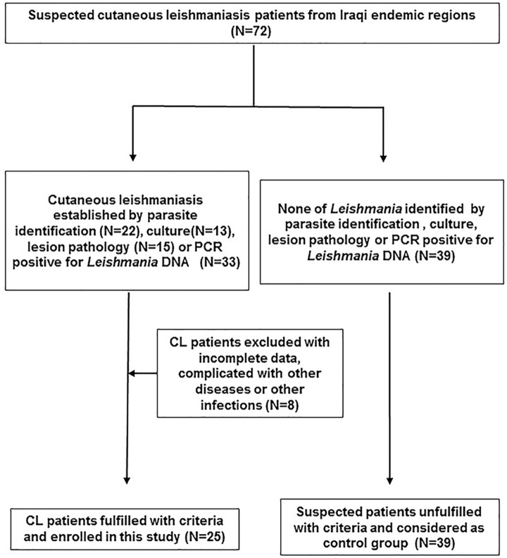
Methods: The definitive diagnosis of CL is based on the parasite identification by microscopic examination directly on lesion smear or parasite culture, PCR amplification of Leishmania-specific internal transcribed spacer 1 (ITS-1). The phylogenetic analysis, the immunopathological examination and the cytokine detection were proceeded after the diagnosis.
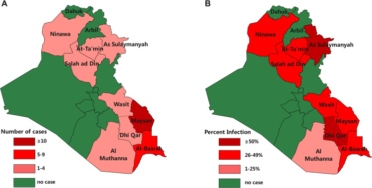
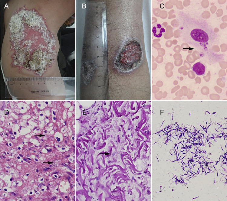
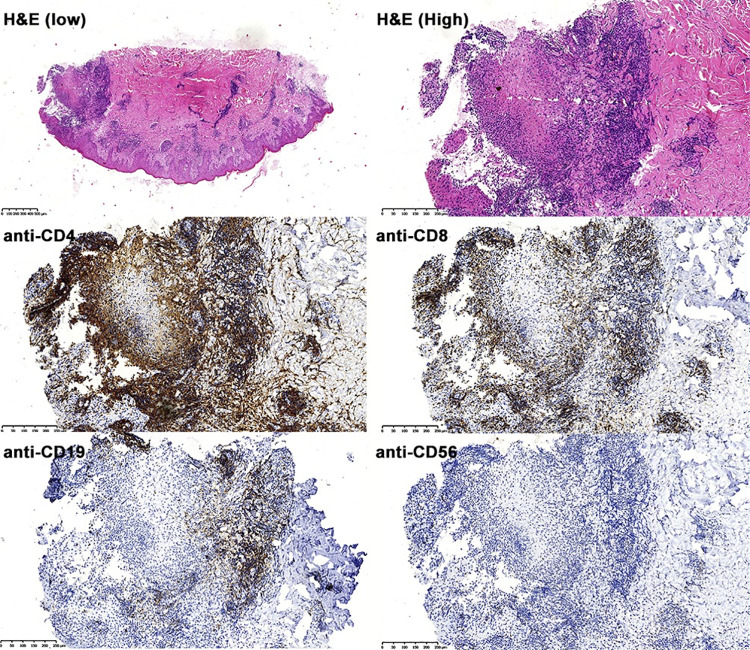
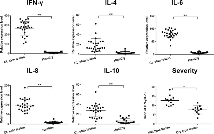
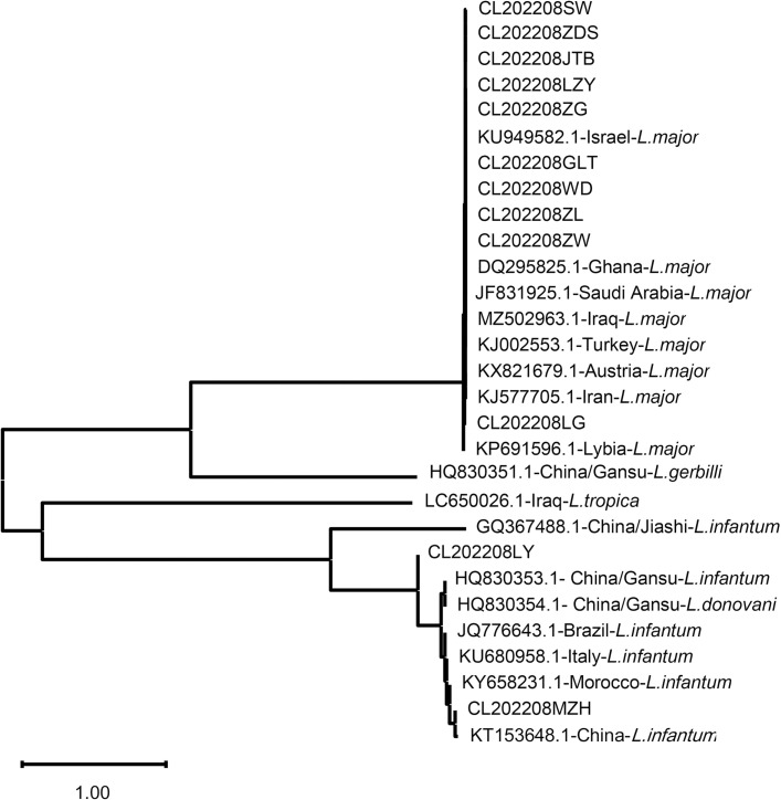
Results: We have identified 25 CL cases in migrant Chinese workers returned from Iraq for the first time with L. major as the major species of infected Leishmania parasite. Clinical features of the Iraq-imported CL include the history of skin exposure to sandflies bite and the lesions mostly on the exposed limbs. More ulcerative wet lesion was observed than nodular dry lesion. PCR is not only used to detect Leishmania parasite with high sensitivity, but also to identify the species of infected parasite through sequencing the amplified Leishmania-specific ITS-1 gene. The phylogenetic analysis based on the amplified ITS-1 sequences revealed that the infected Leishmania was closed related to the species and strains endemic in Iraq. The immunopathological examination revealed the T-cell filtrated cellular immune response with less B cells and NK cells involved. The cytokine profile measured in the skin lesion also confirmed the Th1 cellular response with higher expression levels of IFN-γ, IL-6 and IL-8. The skin lesions in CL patients were healed after being treated locally with antimony.
Conclusions: The clinical and parasitological features of these Chinese CL cases imported from Iraq provide useful information for the diagnosis and treatment of CL that is not commonly seen in Chinese local population.
Copyright: © 2024 Bi et al. This is an open access article distributed under the terms of the Creative Commons Attribution License, which permits unrestricted use, distribution, and reproduction in any medium, provided the original author and source are credited.
Conflict of interest statement
The authors declare that they have no competing interests.
Figures
Similar articles
-
Role of Molecular Diagnosis in Imported Cutaneous Leishmaniasis and Its Public Health Significance in India.Pathogens. 2025 Apr 30;14(5):436. doi: 10.3390/pathogens14050436. Pathogens. 2025. PMID: 40430757 Free PMC article.
-
Treatment outcome of imported cutaneous leishmaniasis among travelers and migrants infected with Leishmania major and Leishmania tropica: a retrospective study in European centers 2013 to 2019.Int J Infect Dis. 2022 Sep;122:375-381. doi: 10.1016/j.ijid.2022.06.025. Epub 2022 Jun 18. Int J Infect Dis. 2022. PMID: 35728749
-
Imported cutaneous leishmaniasis: molecular investigation unveils Leishmania major in Bangladesh.Parasit Vectors. 2019 Nov 7;12(1):527. doi: 10.1186/s13071-019-3771-6. Parasit Vectors. 2019. PMID: 31699125 Free PMC article.
-
Leishmaniasis: recognition and management with a focus on the immunocompromised patient.Am J Clin Dermatol. 2002;3(2):91-105. doi: 10.2165/00128071-200203020-00003. Am J Clin Dermatol. 2002. PMID: 11893221 Review.
-
Phylogenetic analysis of kinetoplast DNA: kDNA of Leishmania tropica in Thi-Qar province, Iraq.Comp Immunol Microbiol Infect Dis. 2021 Oct;78:101696. doi: 10.1016/j.cimid.2021.101696. Epub 2021 Aug 12. Comp Immunol Microbiol Infect Dis. 2021. PMID: 34416483 Review.
Cited by
-
Instrument-Free Point-of-Care Diagnostic for Leishmania Parasites.Diagnostics (Basel). 2024 Dec 5;14(23):2744. doi: 10.3390/diagnostics14232744. Diagnostics (Basel). 2024. PMID: 39682651 Free PMC article.
References
-
- WHO. Leishmaniasis. 2023. www.who.int/news-room/fact-sheets/detail/leishmaniasis
MeSH terms
Substances
LinkOut - more resources
Full Text Sources
