. 2024 Mar 12;121(11):e2307813120.
doi: 10.1073/pnas.2307813120. Epub 2024 Mar 4.

Thiophene-based lipids for mRNA delivery to pulmonary and retinal tissues

Affiliations

Thiophene-based lipids for mRNA delivery to pulmonary and retinal tissues

Yulia Eygeris et al. Proc Natl Acad Sci U S A. .

Abstract

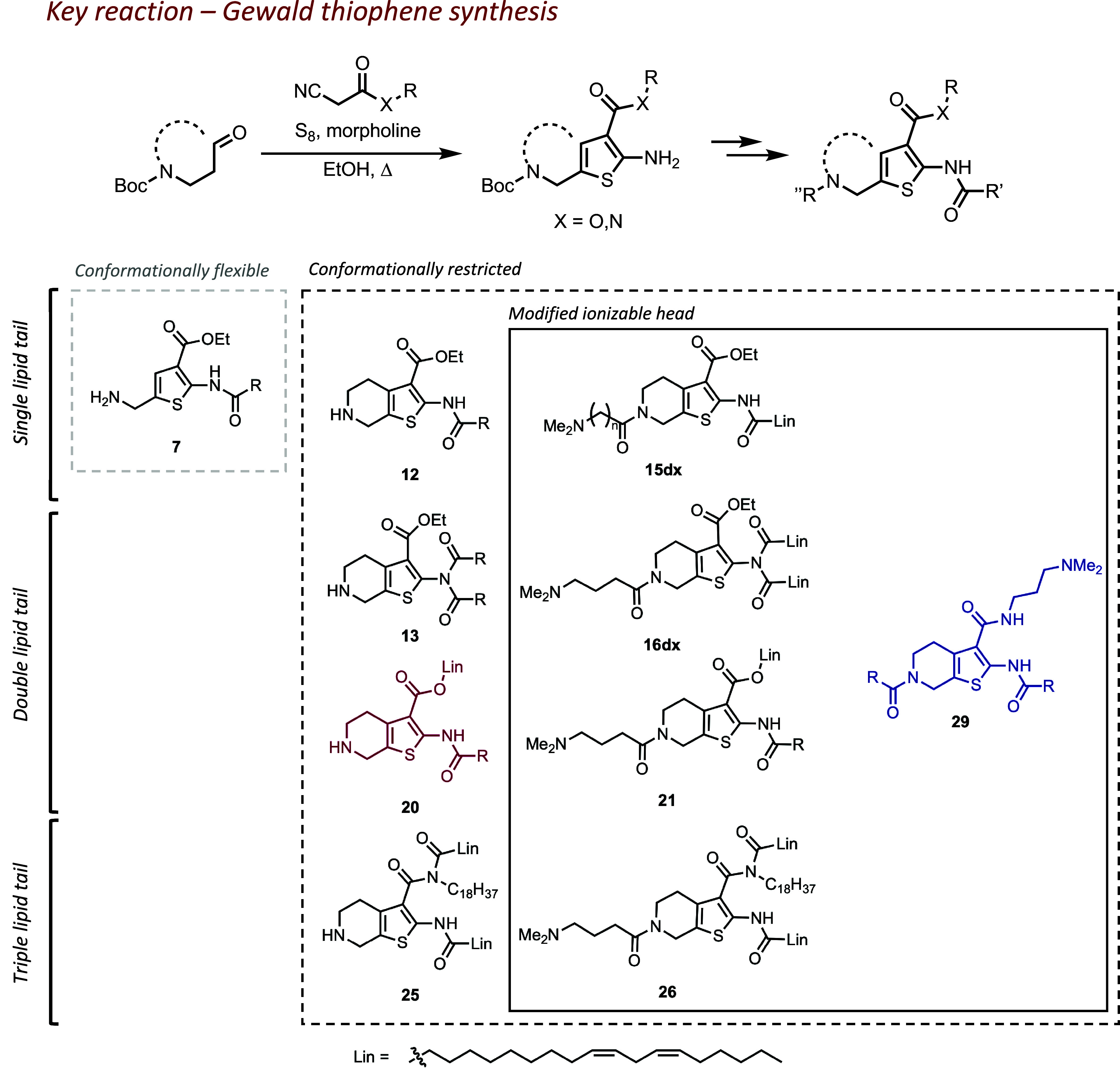
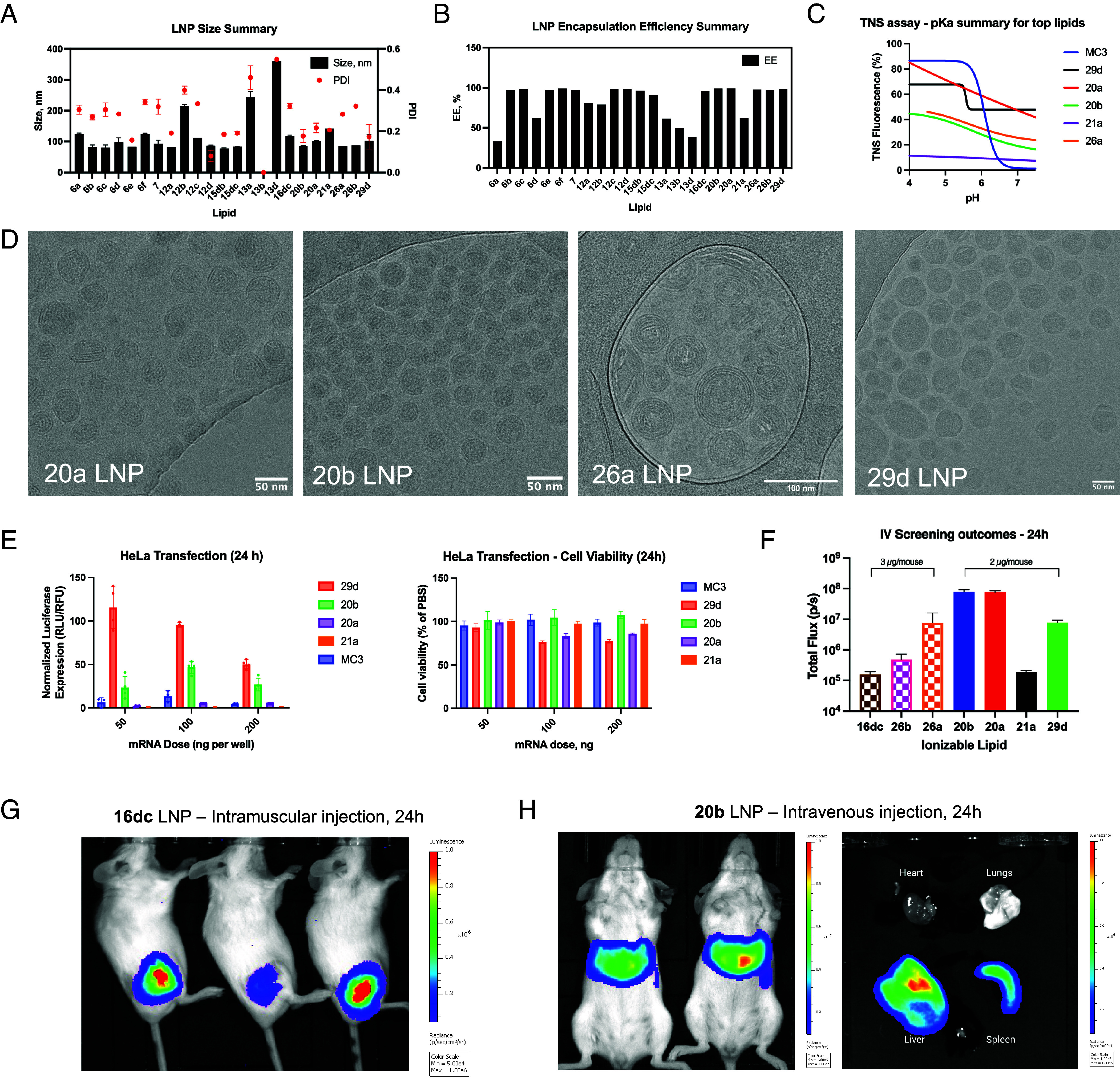
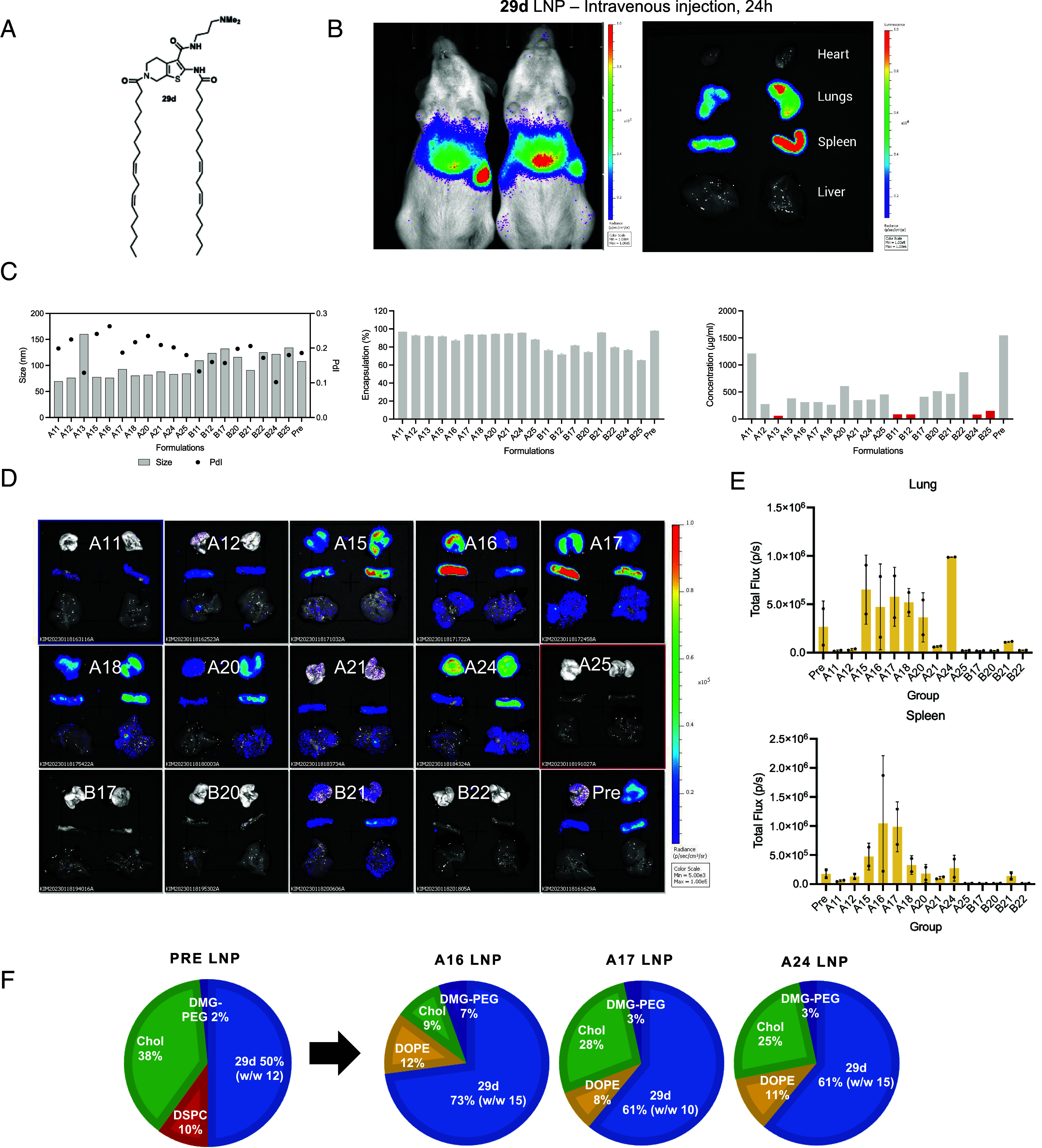
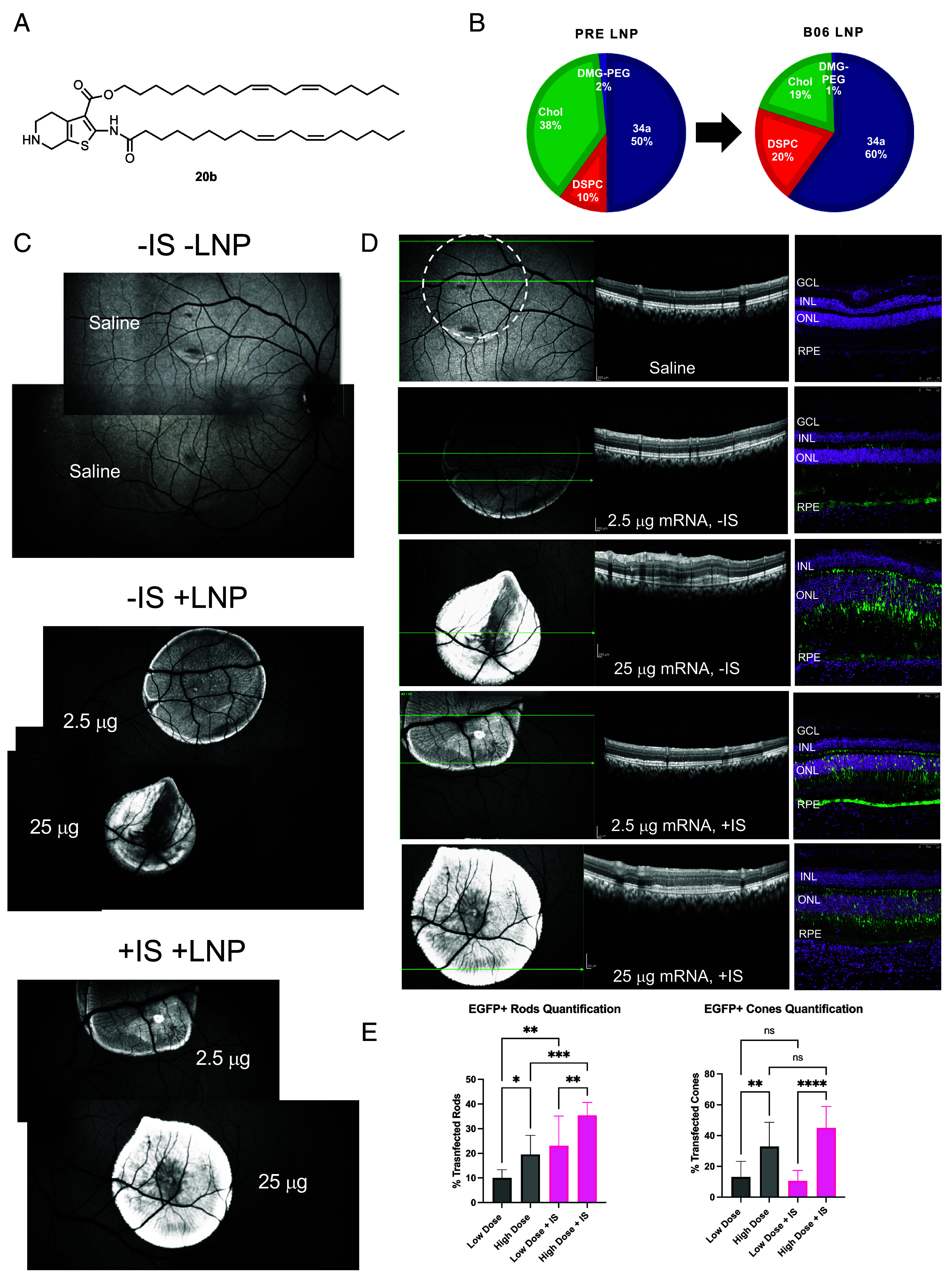
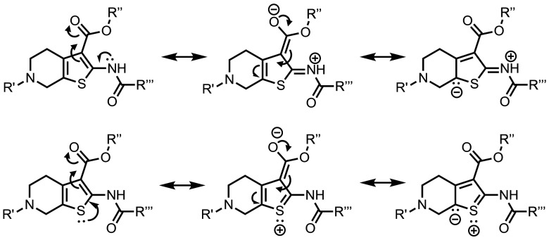
Lipid nanoparticles (LNPs) largely rely on ionizable lipids to yield successful nucleic acid delivery via electrostatic disruption of the endosomal membrane. Here, we report the identification and evaluation of ionizable lipids containing a thiophene moiety (Thio-lipids). The Thio-lipids can be readily synthesized via the Gewald reaction, allowing for modular lipid design with functional constituents at various positions of the thiophene ring. Through the rational design of ionizable lipid structure, we prepared 47 Thio-lipids and identified some structural criteria required in Thio-lipids for efficient mRNA (messenger RNA) encapsulation and delivery in vitro and in vivo. Notably, none of the tested lipids have a pH-response profile like traditional ionizable lipids, potentially due to the electron delocalization in the thiophene core. Placement of the tails and localization of the ionizable headgroup in the thiophene core can endow the nanoparticles with the capability to reach various tissues. Using high-throughput formulation and barcoding techniques, we optimized the formulations to select two top lipids-20b and 29d-and investigated their biodistribution in mice. Lipid 20b enabled LNPs to transfect the liver and spleen, and 29d LNP transfected the lung and spleen. Unexpectedly, LNP with lipid 20b was especially potent in mRNA delivery to the retina with no acute toxicity, leading to the successful delivery to the photoreceptors and retinal pigment epithelium in non-human primates.

Keywords: ionizable lipid; lipid nanoparticle; mRNA delivery.


Conflict of interest statement

Competing interests statement:Y.E., M. Gupta, J.K., M. Gautam, J.R., and G.S. are inventors on a patent application pertinent to this work filed by the Oregon State University. Y.E., D.N., and A.T. have stock options and advisory roles with EnterX Bio. G.S. is a cofounder of EnterX Bio. EnterX Bio has a scientific research agreement with Oregon State University. G.S. has a conflict management plan at Oregon State University. The other authors declare no competing interests.

Figures

Fig. 1.
Fig. 1.
Synthetic evolution of thiophene-based lipids (Lin = linoleic lipid tail) and the scope of the structures discussed in this work. Lipids are grouped by structural criteria: 1) flexible or restricted conformation of the thiophene core; 2) modification of the ionizable head; and 3) number of lipid tails present in a molecule. Compound enumeration begins from precursors; full synthetic information is available in SI Appendix.
Fig. 2.
Fig. 2.
Physicochemical characterization of LNPs: (A) size and PDI of LNPs; (B) mRNA encapsulation efficiency as determined by the modified RiboGreen assay; (C) determination of pKa for select lipids in LNPs via 2-(p-toluidino) naphthalene-6-sulfonic acid (TNS) assay, typical pH-responsiveness of ionizable lipid represented by MC3 LNP; (D) cryoTEM micrographs illustrating LNP morphologies with select lipids; (E) comparison of 29d, 20b, 20a, and 21a LNP efficacy and cell viability in HeLa; (F) Bioluminescence imaging summary for intravenous administration of LNPs, note the dose difference. (G) Representative in vivo bioluminescence images after an IM administration of LNPs incorporating 16dc; (H) Representative in vivo and ex vivo bioluminescence images after an IV administration of LNPs incorporating lipid 20b, the most potent lipid in the tested series.
Fig. 3.
Fig. 3.
29d LNP formulation optimization for improved lung delivery. (A) Structure of lipid 29d; (B) Representative in vivo and ex vivo bioluminescence images after an IV administration of LNPs incorporating lipid 29d, (C) Summary of LNP formulation prescreening includes analysis of nanoparticle size, polydispersity, and mRNA encapsulation and loading efficiency; (D) results of intravenous screening of most robust formulations. Most LNPs showed preferential tropism toward the lung and spleen; the quantification of luminescence in those organs 24 h after IV administration is shown in panel (E). The top formulations A16, A17, and A24 are illustrated in panel (F).
Fig. 4.
Fig. 4.
Evaluation of the top ionizable lipid 20b in the NHP eye after a subretinal administration of EGFP mRNA 20b LNPs. (A) structure of lipid 20b and the formulation used (B). (C) FAF image montage illustrating the relative EGFP intensity in the back of the eye changing in response to the dose. Saline was included as negative control. −IS animal received standard treatment, while +IS animal also received an immunosuppressive treatment (SI Appendix). (C) Representative OCT suggests negligible retinal toxicity for low-dose treatment (the dashed circle indicates the location of the saline bleb; green arrows indicate the scan plane). Significant perturbation was observed in high-dose −IS treatment compared to saline control, less so in high-dose +IS. (D) Representative IF images confirm disruption of retinal morphology in the 25 μg −IS group (DAPI—magenta, EGFP—green; orientation preserved between OCT and IF). (E) image-based quantification of photoreceptor transfection. Stronger EGFP signal correlated with higher dose or IS treatment (N = 1 eye; the statistical analysis is shown for the quantification in different areas of the injection blebs (n = 10). Results are reported as mean and SD after 1-way ANOVA with the Tukey multiple comparison test; *: P 0.0332; **: P 0.0021; ***: P 0.0002; ****: P < 0.0001).
Fig. 5.
Fig. 5.
Example resonance structures of Thio-lipids demonstrating extended electron delocalization. The contribution of resonance forms may explain the broad ionization profile of Thio-lipids since heteroatoms (e.g., nitrogen and sulfur) may carry a partial positive charge.

References

    1. Hou X., Zaks T., Langer R., Dong Y., Lipid nanoparticles for mRNA delivery. Nat. Rev. Mater 6, 1078–1094 (2021). - PMC - PubMed
    1. Zhang Y., Sun C., Wang C., Jankovic K. E., Dong Y., Lipids and lipid derivatives for RNA delivery. Chem. Rev. 121, 12181–12277 (2021). - PMC - PubMed
    1. Cheng Q., et al. , Selective organ targeting (SORT) nanoparticles for tissue-specific mRNA delivery and CRISPR–Cas gene editing. Nat. Nanotechnol. 15, 313–320 (2020). - PMC - PubMed
    1. Dilliard S. A., Cheng Q., Siegwart D. J., On the mechanism of tissue-specific mRNA delivery by selective organ targeting nanoparticles. Proc. Natl. Acad. Sci. U.S.A. 118, e2109256118 (2021). - PMC - PubMed
    1. Qiu M., et al. , Lung-selective mRNA delivery of synthetic lipid nanoparticles for the treatment of pulmonary lymphangioleiomyomatosis. Proc. Natl. Acad. Sci. U.S.A. 119, e2116271119 (2022). - PMC - PubMed

LinkOut - more resources