. 2024 Mar 19;63(6):777-787.
doi: 10.1021/acs.biochem.3c00636. Epub 2024 Mar 4.

Zα Domain of ADAR1 Binds to an A-Form-like Nucleic Acid Duplex with Low Micromolar Affinity

Affiliations

Zα Domain of ADAR1 Binds to an A-Form-like Nucleic Acid Duplex with Low Micromolar Affinity

Parker J Nichols et al. Biochemistry. .

Abstract

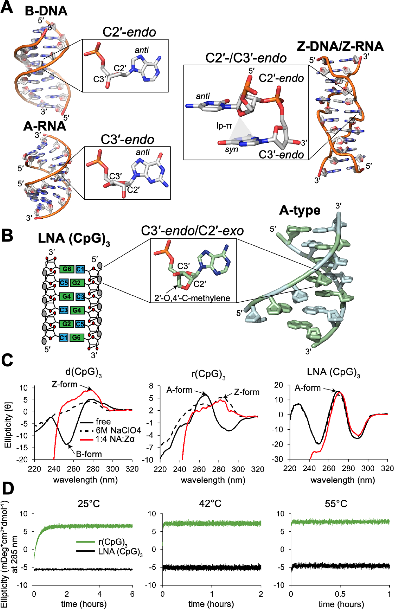
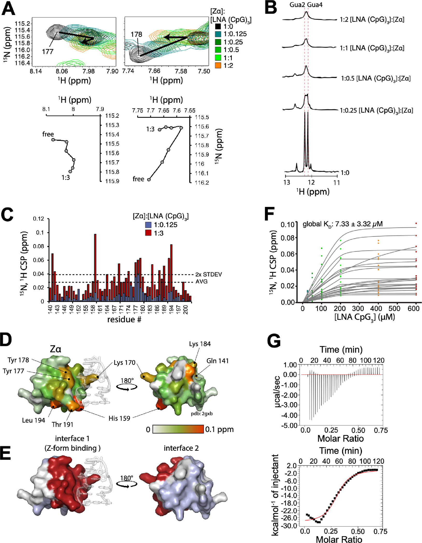
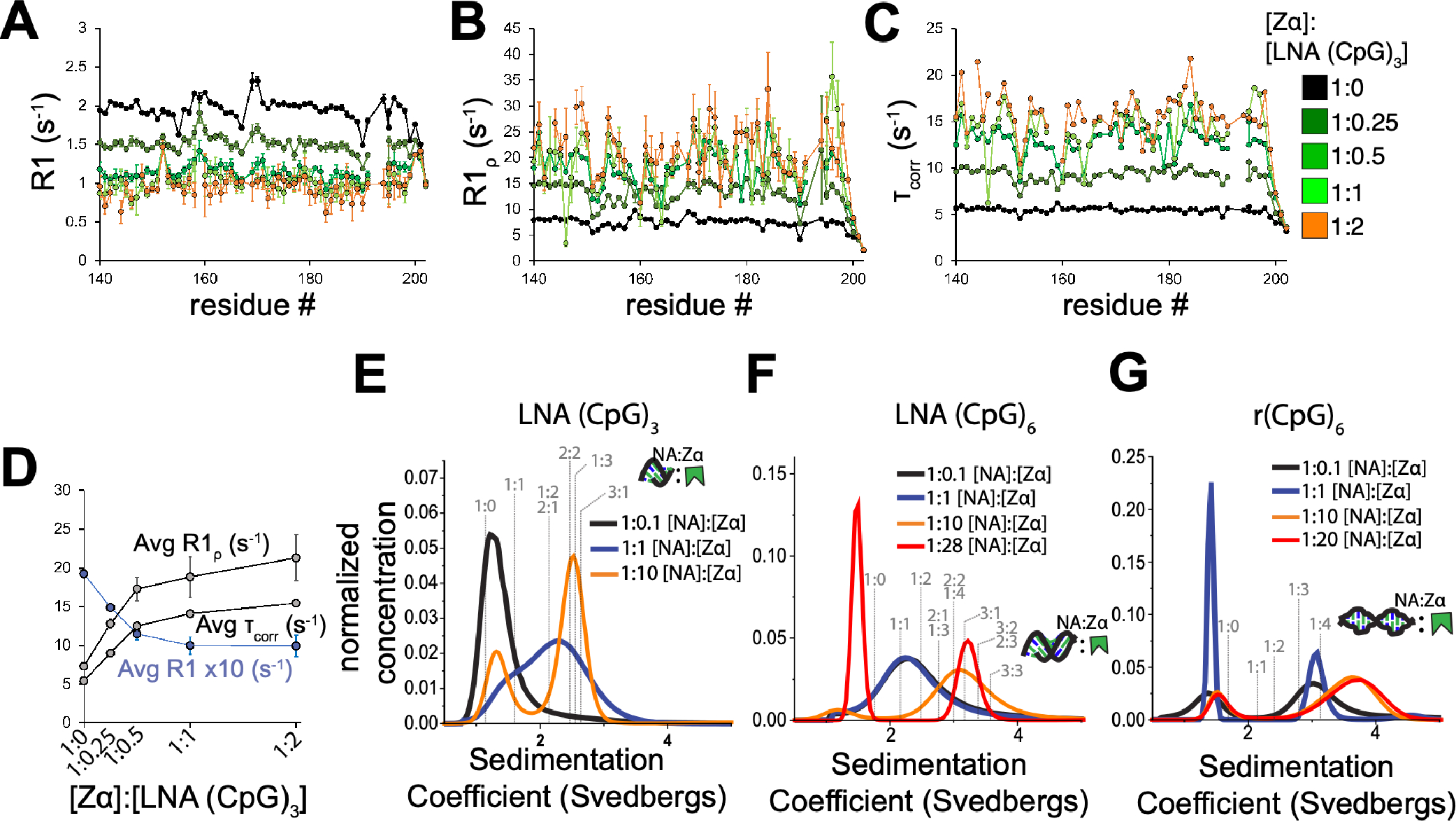
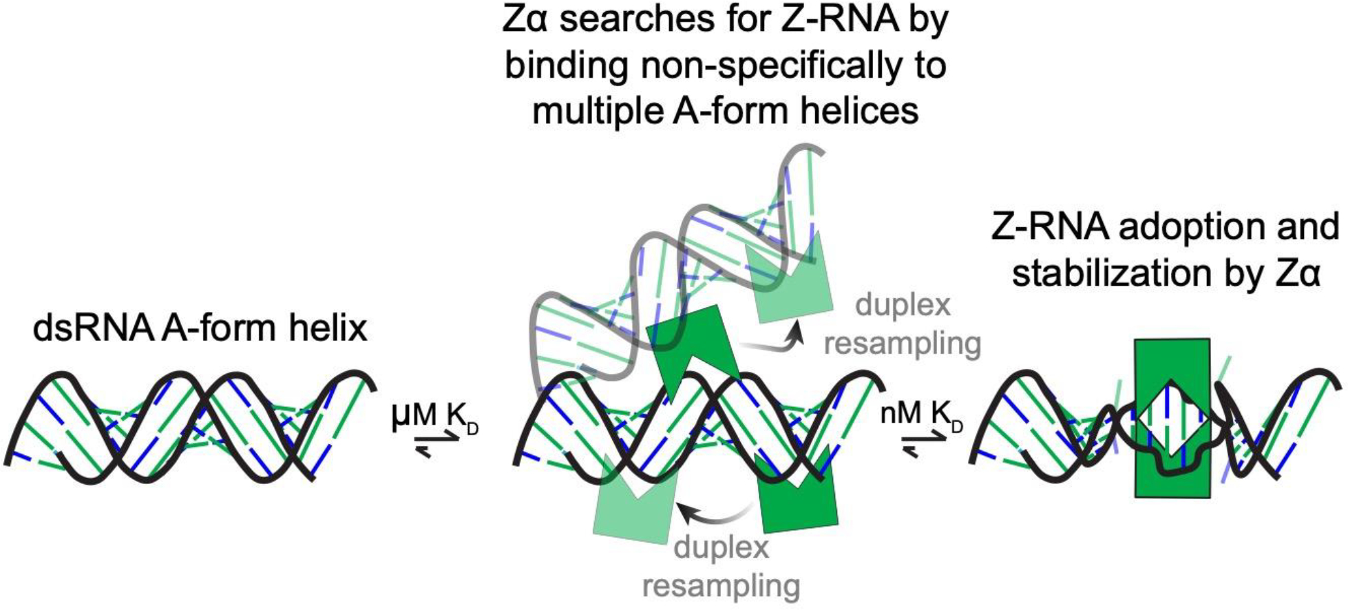
The left-handed Z-conformation of nucleic acids can be adopted by both DNA and RNA when bound by Zα domains found within a variety of viral and innate immune response proteins. While Z-form adoption is preferred by certain sequences, such as the commonly studied (CpG)n repeats, Zα has been reported to bind to a wide range of sequence contexts. Studying how Zα interacts with B-/A-form helices prior to their conversion to the Z-conformation is challenging as binding coincides with Z-form adoption. Here, we studied the binding of Zα fromHomo sapiens ADAR1 to a locked "A-type" version of the (CpG)3 construct (LNA (CpG)3) where the sugar pucker is locked into the C3'-endo/C2'-exo conformation, which prevents the duplex from adopting the alternating C2'/C3'-endo sugar puckers found in the Z-conformation. Using NMR and other biophysical techniques, we find that ZαADAR1 binds to the LNA (CpG)3 using a similar interface as for Z-form binding, with a dissociation constant (KD) of ∼4 μM. In contrast to Z-DNA/Z-RNA, where two ZαADAR1 bind to every 6 bp stretch, our data suggests that ZαADAR1 binds to multiple LNA molecules, indicating a completely different binding mode. Because ZαADAR1 binds relatively tightly to a non-Z-form model, its binding to B/A-form helices may need to be considered when experiments are carried out which attempt to identify the Z-form targets of Zα domains. The use of LNA constructs may be beneficial in experiments where negative controls for Z-form adoption are needed.


Figures

Figure 1.
Figure 1.
A locked version of the (CpG)3 duplex does not adopt the Z-form conformation by high-salt or in the presence of Zα. (a) Structural depictions of a B-form helix and its C2′-endo sugar pucker, an A-form helix and corresponding C3′-endo sugar pucker, and a Z-form helix and its alternating C2′-endo/anti C3′-endo/syn configuration (from 5′ to 3’). 3D depictions were made using PDB 6CQ3 (B-form helix), 2JXQ (A-form helix), and 4OCB (Z-form helix). (b) 2D cartoon schematic of the locked (CpG)3 construct (left) and 3D structure of a fully locked duplex (right, using PDB: 2X2Q). The methylene bridge which locks the sugar pucker into C3’-endo/C2’-exo and thus the duplex into an A-type conformation is highlighted. (c) CD spectra from wavelengths of 220–320 nm of the d(CpG)3, r(CpG)3, and LNA (CpG)3 duplexes in low salt (free, black solid lines), high-salt (6M NaClO4, slashed black lines), and in complex with Zα (1:4 molar ratio of Nucleic Acid:Zα, red lines). The B-form, A-form, and Z-form spectral profiles are indicated. (d) Plots of the ellipticity at 285 nm measured by circular dichroism as a function of time after the addition of saturating amounts of Zα for the r(CpG)3 and LNA (CpG)3 duplexes. The temperature that the process was measured at is indicated at the top of the graphs.
Figure 2.
Figure 2.
ADAR1 binds to the LNA (CpG)3 using a similar interface as for Z-form nucleic acids and with low micromolar affinity. (a) Zoomed in insets of residues Tyr 177 and Ser 178 from the 2D 15N-HSQC titration of LNA (CpG)3 into 15N-labeled ZαADAR1 (full spectra shown in Figure S2). The molar ratio of ZαADAR1:LNA (CpG)3 is color-coded. Below the spectra are graphs plotting the 1H and 15N chemical shifts (ppm) of the same residues throughout the titration. (b) 1D 1H imino spectra following titration of ZαADAR1 into the LNA (CpG)3 construct. The ratio of LNA (CpG)3:ZαADAR1 is indicated on the right-hand side of the spectra. The imino assignment is indicated. (c) Chemical shift perturbations (CSPs) are plotted vs the residue number, and color coded according to the ratio of ZαADAR1:LNA (CpG)3. (d) CSPs from (c) plotted onto the structure of ZαADAR1 bound to the r(CpG)3 duplex in the Z-conformation (PDB: 2GXB). (e) The two interfaces revealed by clustering of CSPs (red is the Z-RNA binding interface, blue a second interface not known to be involved in nucleic acid binding). (f) Global fit of the dissociation constant (KD) from the CSPs as a function of the LNA (CpG)3 concentration. (g) Isothermal calorimetry thermogram of the LNA (CpG)3ADAR1 interaction. Extracted thermodynamic parameters are found in Table 1.
Figure 3.
Figure 3.
The complex stoichiometry of LNA:ZαADAR1 binding is similar to that of Z-RNA. (a) Longitudinal relaxation rates R1, which are sensitive to motions on the ps-ns timescale are plotted on the y-axis vs ZαADAR1’s residue number on the x-axis as a function of increasing LNA (CpG)3 concentration (color-coded, as shown to the right-hand side of (c)). (b) Rotating frame relaxation rates R, which are sensitive to potential slow motions on the μs-ms timescale in addition to the fast motion. (c) Residue-specific tumbling times τcorr calculated from the ratio of R2/R1 (see methods for details). (d) Averages of the corresponding residue-specific R1, R, and τcorr values from (a, b, and c) are plotted versus the molar ratio of ZαADAR1:LNA in the experiment. Measured values are from one set of relaxation rate experiments. Sedimentation coefficient distributions for the LNA (CpG)3 (e), LNA (CpG)6 (f), and r(CpG)6 (g) at decreasing ratios of nucleic acid (NA) to ZαADAR1 as obtained from analytical ultracentrifugation (AUC). Predicted sedimentation coefficients for different theoretical nucleic acid ZαADAR1 complex stoichiometries are shown overlayed on top of the plots. Some of the predicted sedimentation coefficients are identical and are thus overlapped (e.g., for the LNA (CpG)6, 2:2 and 1:4 had the sample predicted sedimentation coefficient).
Figure 4.
Figure 4.
Model incorporating binding of Zα to an A-form helix prior to Z-RNA adoption and stabilization. Zα initially binds to multiple A-form helices with low micromolar affinity and has a fast exchange rate between the free and bound states. This is likely accompanied shuffling between the different duplexes brought into proximity. The bound RNA then transitions to the Z-conformation which is recognized by the Zα domains leading to a significantly lower dissociation constant and a slow off-rate, thus stabilizing the Z-RNA conformation.

Similar articles

References

    1. Wolfram S Principles of Nucleic Acid Structure, 1st ed.; Springer New York: New York, 1984.
    1. Plavee J; Tong W; Chattopadhyaya J How Do the Gauche and Anomeric Effects Drive the Pseudorotational Equilibrium of the Pentofuranose Moiety of Nucleosides. J. Am. Chem. Soc. 1993. 10.1021/ja00074a046. - DOI
    1. Plavec J; Thibaudeau C; Chattopadhyaya J How Does the 2’-Hydroxy Group Drive the Pseudorotational Equilibrium in Nucleoside and Nucleotide by the Tuning of the 3’-Gauche Effect? J. Am. Chem. Soc. 1994. 10.1021/ja00094a009. - DOI
    1. Hall K; Cruz P; Tinoco I; Jovin TM; Van De Sande JH “Z-RNA” - A Left-Handed RNA Double Helix. Nature 1984. 10.1038/311584a0. - DOI - PubMed
    1. Schwartz T; Rould MA; Lowenhaupt K; Herbert A; Rich A Crystal Structure of the Zα Domain of the Human Editing Enzyme ADAR1 Bound to Left-Handed Z-DNA. Science (80-. ). 1999. 10.1126/science.284.5421.1841. - DOI - PubMed

Publication types