

Risk factors and implications associated with ultrasound-diagnosed nephrocalcinosis in cats with chronic kidney disease
- PMID: 38438128
- PMCID: PMC11099775
- DOI: 10.1111/jvim.17034
Risk factors and implications associated with ultrasound-diagnosed nephrocalcinosis in cats with chronic kidney disease
Abstract
Background: Microscopic nephrocalcinosis is a common pathological feature of chronic kidney disease (CKD) in cats. Detection of macroscopic nephrocalcinosis using ultrasonography and its implications remain unexplored.
Objectives: Identify risk factors associated with ultrasound-diagnosed nephrocalcinosis and evaluate the influence of nephrocalcinosis on CKD progression.
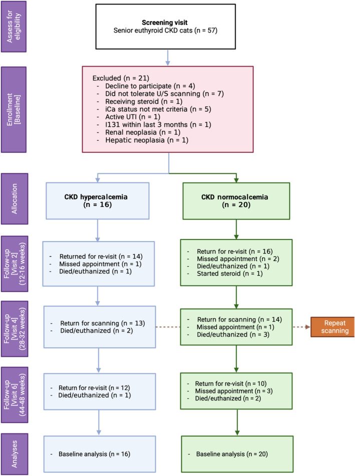
Animals: Thirty-six euthyroid client-owned cats with CKD.
Methods: Prospective cohort study. Cats with CKD with and without ionized hypercalcemia were enrolled for renal ultrasonography. Cats were categorized according to the presence or absence of ultrasound-diagnosed nephrocalcinosis. Binary logistic regression was performed to identify nephrocalcinosis risk factors. The influence of nephrocalcinosis on CKD progression was assessed using linear mixed models.
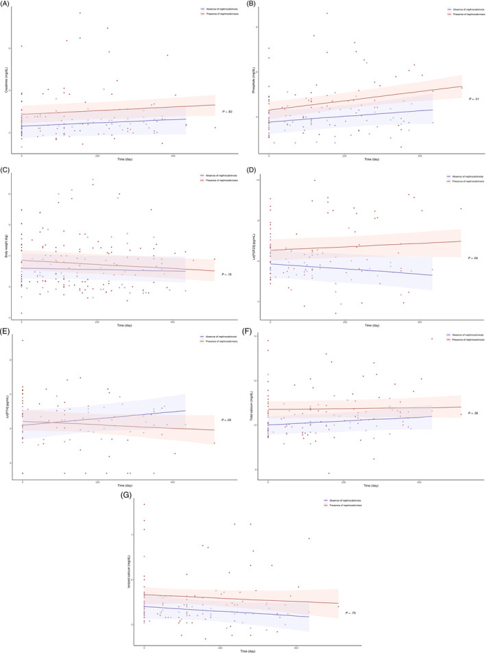
Results: Ultrasound-diagnosed nephrocalcinosis was evident in 61% of CKD cats overall, with increased prevalence (81%) in those with hypercalcemia. At enrollment, higher blood ionized calcium concentration (odds ratio [OR], 1.27 per 0.1 mg/dL; P = .01), plasma phosphate concentration (OR, 1.16 per 0.1 mg/dL; P = .05), plasma creatinine concentration (OR, 1.29 per 0.1 mg/dL; P = .02) and alanine aminotransferase activity (OR, 2.08 per 10 U/L; P = .04) were independent nephrocalcinosis risk factors. The rate of change in log-transformed fibroblast growth factor-23 differed significantly between groups (P = .04). Cats with CKD and nephrocalcinosis had increasing plasma creatinine concentrations (.03 ± .01 mg/dL/month; P = .04) and phosphate concentrations (.06 ± .02 mg/dL/month; P < .001) and decreasing body weight (.02 ± .01 kg/month; P < .001) over time.
Conclusions and clinical importance: Nephrocalcinosis is prevalent in cats with CKD, especially in those with hypercalcemia. This pathological feature appears to be associated with CKD progression in cats.
Keywords: CKD‐MBD; feline; hypercalcemia; mineralization; nephrolithiasis; radiology and diagnostic imaging; ultrasonography.
© 2024 The Authors. Journal of Veterinary Internal Medicine published by Wiley Periodicals LLC on behalf of American College of Veterinary Internal Medicine.
Conflict of interest statement
Pak Kan Tang received a PhD studentship funded by Royal Canin SAS. Rebecca Geddes received funding from Petplan, Royal Canin, an RVC Internal Grant, The Academy of Medical Sciences and The Everycat Foundation; has previously had a consultancy agreement with Boehringer Ingelheim; has received speaking honoraria from Boehringer Ingelheim, Idexx and Royal Canin. Rosanne Jepson received funding from PetPlan, Feline Foundation for Renal Research, RVC Internal Grant, PetSavers, and consultancy agreements: Boehringer Ingelheim, Merial, CEVA. Speaking honoraria: Boehringer Ingelheim, Hills Pet Nutrition, CEVA. Jonathan Elliott has Consultancy agreements with: Elanco Ltd, CEVA Animal Health Ltd, Boehringer Ingelheim Ltd, MSD Animal Health Ltd., Orion Incorp, Idexx Ltd, Waltham Petcare Science Institute, Invetx Inc and Zoetis Ltd. received grant funding from Elanco Ltd, Waltham Centre for Pet Nutrition, Royal Canin SAS, Idexx Ltd., CEVA Animal Health. He is a member of the International Renal Interest Society which receives sponsorship from Zoetis.
Figures
Similar articles
-
Risk factors and implications associated with renal mineralization in chronic kidney disease in cats.J Vet Intern Med. 2022 Mar;36(2):634-646. doi: 10.1111/jvim.16363. Epub 2022 Jan 19. J Vet Intern Med. 2022. PMID: 35043997 Free PMC article.
-
Dietary magnesium supplementation in cats with chronic kidney disease: A prospective double-blind randomized controlled trial.J Vet Intern Med. 2024 Jul-Aug;38(4):2180-2195. doi: 10.1111/jvim.17134. Epub 2024 Jul 1. J Vet Intern Med. 2024. PMID: 38952053 Free PMC article.
-
Detection of nephrocalcinosis using ultrasonography, micro-computed tomography, and histopathology in cats.J Vet Intern Med. 2024 May-Jun;38(3):1553-1562. doi: 10.1111/jvim.17011. Epub 2024 Feb 13. J Vet Intern Med. 2024. PMID: 38348812 Free PMC article.
-
A feline-focused review of chronic kidney disease-mineral and bone disorders - Part 2: Pathophysiology of calcium disorder and extraosseous calcification.Vet J. 2021 Sep;275:105718. doi: 10.1016/j.tvjl.2021.105718. Epub 2021 Jul 27. Vet J. 2021. PMID: 34329743 Review.
-
Feline CKD: Pathophysiology and risk factors--what do we know?J Feline Med Surg. 2013 Sep;15 Suppl 1(1 Suppl):3-14. doi: 10.1177/1098612X13495234. J Feline Med Surg. 2013. PMID: 23999182 Free PMC article. Review.
Cited by
-
Expression of osteogenic proteins in kidneys of cats with nephrocalcinosis.J Vet Intern Med. 2025 Jan-Feb;39(1):e17278. doi: 10.1111/jvim.17278. J Vet Intern Med. 2025. PMID: 39757788 Free PMC article.
References
-
- Evenepoel P, Daenen K, Bammens B, et al. Microscopic nephrocalcinosis in chronic kidney disease patients. Nephrol Dial Transplant. 2015;30(5):843‐848. - PubMed
MeSH terms
Grants and funding
LinkOut - more resources
Full Text Sources
Medical
Miscellaneous
