Review
. 2024 Mar 4;34(3):363-378.
doi: 10.1136/ijgc-2023-004609.

Staging by imaging in gynecologic cancer and the role of ultrasound: an update of European joint consensus statements

Affiliations
Review

Staging by imaging in gynecologic cancer and the role of ultrasound: an update of European joint consensus statements

Daniela Fischerova et al. Int J Gynecol Cancer. .

Abstract

In recent years the role of diagnostic imaging by pelvic ultrasound in the diagnosis and staging of gynecological cancers has been growing exponentially. Evidence from recent prospective multicenter studies has demonstrated high accuracy for pre-operative locoregional ultrasound staging in gynecological cancers. Therefore, in many leading gynecologic oncology units, ultrasound is implemented next to pelvic MRI as the first-line imaging modality for gynecological cancer. The work herein is a consensus statement on the role of pre-operative imaging by ultrasound and other imaging modalities in gynecological cancer, following European Society guidelines.

Keywords: cervical cancer; cross-sectional studies; ovarian cancer; uterine cancer; vulvar and vaginal cancer.


Conflict of interest statement

Competing interests: None declared.

Figures

Figure 1
Figure 1
Ultrasound approaches for gynecological cancer staging: transvaginal (A) and transperineal in combination with transrectal (B) approach for local staging; transabdominal approach using a convex array probe for evaluation of abdominal infiltrated visceral and retroperitoneal lymph nodes, peritoneal, or parenchymal metastases (C); transcutaneous approach with linear array probe for evaluation of inguinofemoral lymph nodes (D).
Figure 2
Figure 2
Imaging by ultrasound, CECT, and FDG-PET-CT (depicting locoregional spread and distant spread in a patient in her 80s, diagnosed with squamous cell vulvar cancer FIGO stage IVB. For local staging, transperineal ultrasound using a convex array probe in the transverse plane allows visualization of tumor infiltration in the clitoris (A) and labia majora bilaterally (B). The same vulvar pathology is depicted as hyperdense tissue on CECT (C) with high FDG-avidity on FDG-PET-CT (D). Regional lymph nodes in the groins are evaluated by transcutaneous ultrasound using a linear array probe (according to the Vulvar International Tumor Analysis (VITA) Group consensus for the evaluation of inguinofemoral lymph nodes) (E, F). Two pathologic lymph nodes (Ln1 and Ln2) above the fascia lata and the femoral vessels on the right side (E, F) are seen: Ln1 is partially infiltrated while Ln2 shows complete infiltration. The same pathology is depicted as enlarged inguinal lymph nodes on CECT (G), highly FDG-avid on FDG-PET-CT (H). Pelvic lymph nodes are assessable by ultrasound using an endoluminal probe inserted transvaginally and metastatic lymph nodes are seen around the right (Ln3) and left (Ln4) (I, J) iliac vessels. The enlarged iliac lymph node is clearly visualized on the right side (Ln3) on CECT (K) and as highly FDG-avid on FDG-PET-CT (L). The location and size of this vulvar lesion made a transvaginal ultrasound approach possible. Online Supplemental Video S1 shows ultrasound and other imaging methods in vulvar cancer. CECT, contrast-enhanced CT; FDG-PET-CT, 18F-fluorodeoxyglucose positron emission tomography combined with CECT. FIGO, International Federation of Gynecology and Obstetrics; Ln, lymph node.
Figure 3
Figure 3
Ultrasound, CECT and FDG-PET-CT in woman in her 70 s diagnosed with squamous cell vaginal cancer FIGO stage III. Transrectal ultrasound depicts a hypoechogenic to isoechogenic, richly vascularized tumor in the lower part of the anterior vaginal wall (A, B); corresponding pathologic mass depicted on axial CECT (C) is slightly contrast-enhancing and is highly FDG-avid on 18FFDG-PET-CT (D).Right and left hypoechogenic enlarged metastatic inguinal lymph nodes (Ln1 and Ln2) were seen on ultrasound (E, F), axial CECT (G), and as FDG-avid lesions on FDG-PET-CT (H). Right and left external iliac lymph nodes (Ln3 and Ln4) demonstrated similar appearance to the involved inguinal lymph nodes on transrectal ultrasound (I, J), axial CECT (K), and FDG-PET-CT (L), indicating metastatic lymph nodes. CECT, contrast-enhanced CT; FDG-PET-CT, 18F-fluorodeoxyglucose positron emission tomography combined with CECT; FIGO, International Federation of Gynecology and Obstetrics; ln, lymph nodes. The Online Supplemental Video S2 shows this tumor in the lower part of the vagina.
Figure 4
Figure 4
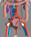
Pre-operative assessment of the disease extent and prognostic factors necessary for planning treatment. The size and tumor topography within the cervix (1), the parametrial spread (ventral, lateral, dorsal; right and left) (2) with the possible involvement of the urinary tract (3), the presence of metastatic pelvic (4), and para-aortic lymph nodes (5) and others. Pre-operative staging shows Online Supplemental Video S3 (available here).
Figure 5
Figure 5
Cervical cancer on the anterior lip of the cervix (A) visualized by MRI (C) and transvaginal ultrasound C,D) in a patient in her 30s diagnosed with squamous cell carcinoma of the cervix, FIGO stage IB3 on radiologic staging. A bulky tumor infiltrating the anterior lip of the cervix and cervical stroma up to the internal os (A) is depicted as hyperintense on T2-weighted MRI (B) and hypoechoic on transvaginal ultrasound (C) with high vascularity depicted by color Doppler (D). Visualization of the tumor vessels supported the drawing of the tumor boundaries (yellow dotted line) and measurements of tumor size (B, D). Bulky cervical tumor clearly visible in the hysterectomy specimen (upper panel) and after transverse sectioning (lower panel) of the tumor at the level of the section line (indicated in A-C) (E). FIGO, International Federation of Gynecology and Obstetrics; MRI, magnetic resonance imaging; TVS, transvaginal scan. The Online Supplemental Video S4 shows an ultrasound of a cervical cancer on the anterior lip.
Figure 6
Figure 6
Synchronous endometrial and ovarian low-grade endometrioid cancer FIGO stage IA3 in a woman in her 30s who underwent transvaginal ultrasound, CECT, and FDG-PET-CT pre-operatively. Uterus with residual low-grade endometrial endometrioid cancer after hysteroscopic resection, with an intrauterine device in situ (A). Left ovary with unilocular-solid tumor histologically verified as low-grade endometrioid carcinoma; of note are papillary projections with smooth rounded contours, high perfusion on color Doppler and intracystic fluid with ground glass echogenicity (B). Pathologic lesion in the left ovary depicted on CECT (C) with contrast-enhancing solid components, and on PET-CT (D) with high FDG-avidity in the solid components; macroscopic appearance of the left ovary after oophorectomy (E, F). CECT, contrast-enhanced CT; FDG-PET-CT, 18F-fluorodeoxyglucose positron emission tomography combined with CECT. FIGO, International Federation of Gynecology and Obstetrics.The Online Supplemental Video S5 shows this case.
Figure 7
Figure 7
Transvaginal ultrasound (A–D), pelvic MRI (E–H), and abdominal CECT (I–L) in a woman in her 50 s diagnosed with clear cell endometrial cancer FIGO stage IIIA1. Transvaginal ultrasound depicts an isoechogenic uterine tumor infiltrating more than 50% of the myometrial wall (A); with protrusion but no invasion into the proximal endocervix, the distance from the external cervical os to the lower margin of the tumor is 22 mm (B); color Doppler depicts a moderately vascularized solid tumor of the right ovary (C); and a multilocular-solid tumor in the left ovary with moderately vascularized solid components (D). Pelvic MRI with T2-weighted sagittal (E) and T2/T1-weighted axial (G, H) series and DWI (high b-value image) (F) depicts a large, hyperintense mass in the uterus extending from the uterine fundus down to the cervix (E). The tumor exhibits restricted diffusion depicted as hyperintensity on the DWI image (F) with low apparent diffusion coefficient (ADC) value on the ADC map (not shown), indicating malignant tumor. On axial T2-weighted MRI small cysts are seen in the right ovarian tumor (G) as hyperintense regions in the anterior part. The left ovarian tumor depicted on T1-weighted MRI is hypointense due to cystic portion (H). Abdominal CECT depicts an irregular large uterine mass (I, K) and the right (I, K) and left (J, L) ovarian tumors with cystic lesions and contrast-enhancing septae. On axial CT at the level of the uterus (K) and at the level above the uterus (L) hypodense areas in the left ovary (L) and the right ovarian tumors (K) indicating cystic spaces. DWI, diffusion-weighted imaging; CECT, contrast-enhanced CT; FIGO, International Federation of Gynecology and Obstetrics. Online Supplemental Video S6 shows this case of endometrial cancer.
Figure 8
Figure 8
Characteristic imaging findings on abdominal ultrasound, whole body (WB)-MRI (showing the abdomen) with DWI and at laparoscopy in a woman in her 50 s diagnosed with FIGO Stage IVB high-grade serous carcinoma of the ovary. Abdominal convex array and transvaginal ultrasound (first column), axial CE-T1-weighted fat-suppressed (CE-T1WI-FS) MRI (part of WB-MRI) (second column), CE-T1WI-FS fused with DWI (high b-value images) (third column) and laparoscopy findings (fourth column). The imaging findings confirmed by laparoscopy indicate visceral hepatic carcinomatosis marked with arrows on the surface of the liver (A–D), mainly cystic carcinomatosis lesions in the lower part of right paracolic gutter (E–H) and large infracolic omental cake (I–L). Peritoneal carcinomatosis is contrast-enhancing (seen as hyperintensity on the WB-MRI series; second column) and exhibits restricted diffusion (seen as hyperintensity on the fused WB-MRI fused with DWI; third column). CE, contrast-enhanced; DWI, diffusion-weighted imaging; FIGO, International Federation of Gynecology and Obstetrics; FS, fat suppression; WB-MRI, whole-body MRI. Online Supplemental Video S8 shows this case of tubo-ovarian cancer spread.

Similar articles

Cited by

References

    1. Andreotti RF, Timmerman D, Strachowski LM, et al. . O-RADS US risk stratification and management system: a consensus guideline from the ACR ovarian-adnexal reporting and data system committee. Radiology 2020;294:168–85. 10.1148/radiol.2019191150 - DOI - PubMed
    1. Van Calster B, Van Hoorde K, Valentin L, et al. . Evaluating the risk of ovarian cancer before surgery using the ADNEX model to differentiate between benign, borderline, early and advanced stage invasive, and secondary metastatic tumours: prospective multicentre diagnostic study. BMJ 2014;349:g5920. 10.1136/bmj.g5920 - DOI - PMC - PubMed
    1. Andreotti RF, Timmerman D, Benacerraf BR, et al. . Ovarian-adnexal reporting lexicon for ultrasound: a white paper of the ACR ovarian-adnexal reporting and data system committee. J Am Coll Radiol 2018;15:1415–29. 10.1016/j.jacr.2018.07.004 - DOI - PubMed
    1. TNM classification of malignant tumours, 8th Edn. 2016.
    1. Eisenhauer EA, Therasse P, Bogaerts J, et al. . New response evaluation criteria in solid tumours: revised RECIST guideline (version 1.1). Eur J Cancer 2009;45:228–47. 10.1016/j.ejca.2008.10.026 - DOI - PubMed

Publication types

LinkOut - more resources