. 2024 Apr 10;44(15):e1728232024.
doi: 10.1523/JNEUROSCI.1728-23.2024.

RANBP17 Overexpression Restores Nucleocytoplasmic Transport and Ameliorates Neurodevelopment in Induced DYT1 Dystonia Motor Neurons

Affiliations

RANBP17 Overexpression Restores Nucleocytoplasmic Transport and Ameliorates Neurodevelopment in Induced DYT1 Dystonia Motor Neurons

Masuma Akter et al. J Neurosci. .

Abstract

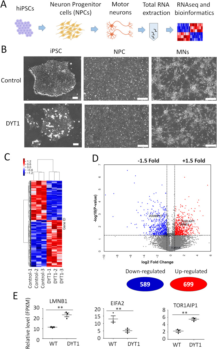
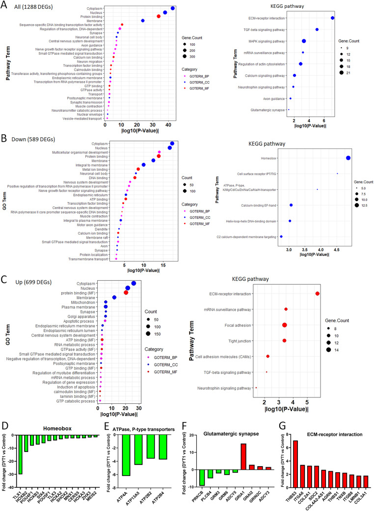
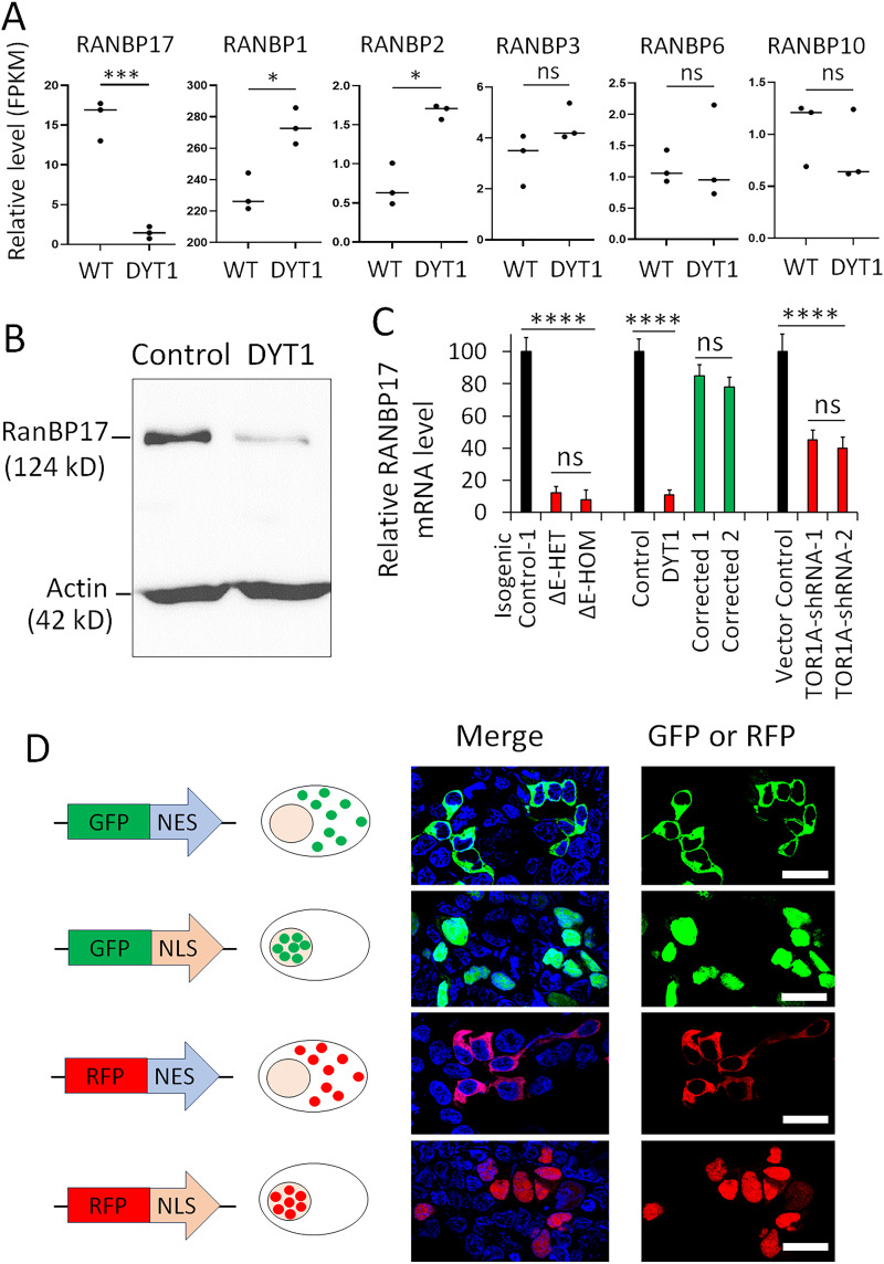
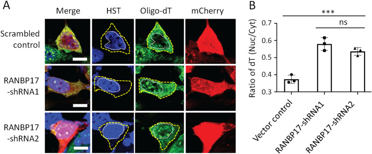
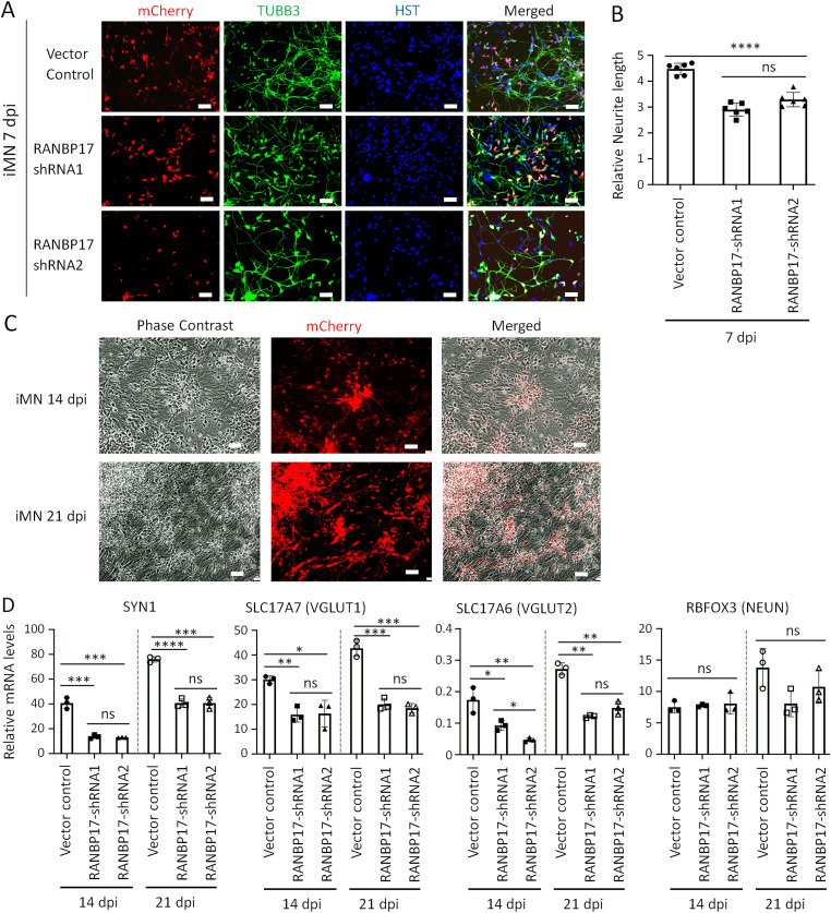
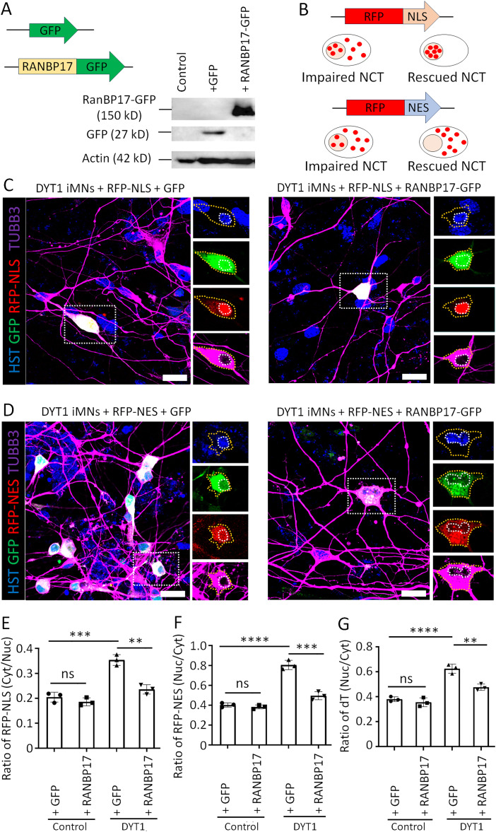
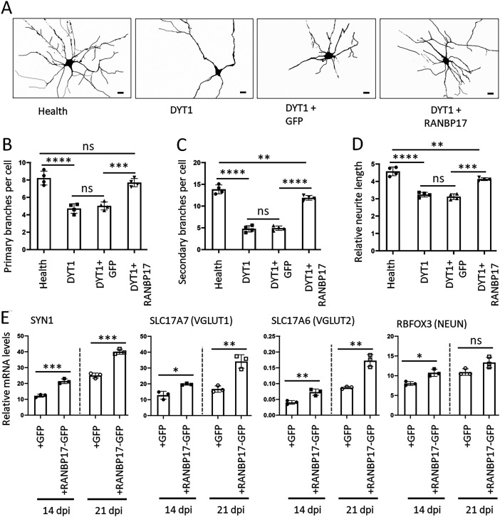
DYT1 dystonia is a debilitating neurological movement disorder, and it represents the most frequent and severe form of hereditary primary dystonia. There is currently no cure for this disease due to its unclear pathogenesis. In our previous study utilizing patient-specific motor neurons (MNs), we identified distinct cellular deficits associated with the disease, including a deformed nucleus, disrupted neurodevelopment, and compromised nucleocytoplasmic transport (NCT) functions. However, the precise molecular mechanisms underlying these cellular impairments have remained elusive. In this study, we revealed the genome-wide changes in gene expression in DYT1 MNs through transcriptomic analysis. We found that those dysregulated genes are intricately involved in neurodevelopment and various biological processes. Interestingly, we identified that the expression level of RANBP17, a RAN-binding protein crucial for NCT regulation, exhibited a significant reduction in DYT1 MNs. By manipulating RANBP17 expression, we further demonstrated that RANBP17 plays an important role in facilitating the nuclear transport of both protein and transcript cargos in induced human neurons. Excitingly, the overexpression of RANBP17 emerged as a substantial mitigating factor, effectively restoring impaired NCT activity and rescuing neurodevelopmental deficits observed in DYT1 MNs. These findings shed light on the intricate molecular underpinnings of impaired NCT in DYT1 neurons and provide novel insights into the pathophysiology of DYT1 dystonia, potentially leading to the development of innovative treatment strategies.

Keywords: RANBP17; Torsin ATPase; dystonia; human-induced pluripotent stem cells (hiPSCs); motor neurons; neurodevelopment; nucleocytoplasmic transport.


Conflict of interest statement

The authors declare no competing financial interests.

Figures

Figure 1.
Figure 1.
Identification of dysregulated genes in human DYT1 MNs. A, Schematic shows the workflow of identification of dysregulated genes from iPSC-derived MNs. B, Representative micrographs of cells at different stages during the generation of human MNs. iPSCs, induced pluripotent stem cells; NPCs, neuronal progenitor cells; and MNs, motor neurons. Scale bar, 100 µm. C, Heat map of RNAseq results of DEGs in DYT1 MNs compared with healthy controls. D, Volcano plot shows the upregulated and downregulated DEGs in DYT1 MNs. Six hundred ninety-nine genes (in red) were upregulated, and 589 genes (in blue) were downregulated. E, Some interesting targets have been identified with disrupted gene expression in DYT1 MNs. FPKM, Fragment Per Kilobase of Million mapped read. **p < 0.01. Student’s t tests.
Figure 2.
Figure 2.
Dysregulated genes in human DYT1 MNs are comprehensively involved in neurodevelopment and various BPs. A, GO analysis of all significant DEGs in DYT1 MNs. Some terms of BP, CC, and MF and KEGG pathways were presented. B, Representative results of GO analysis and KEGG pathways of significantly upregulated genes in DYT1 MNs. C, Representative results of GO analysis and KEGG pathways of significantly downregulated genes in DYT1 MNs. D–G, Some groups of DEGs, including downregulated homeobox genes (D), downregulated ATPases (E), down- and upregulated glutamatergic synapse-related genes (F), and mostly upregulated enriched genes in the KEGG pathway of the ECM–receptor interaction (G).
Figure 3.
Figure 3.
RANBP17 expression decreased in DYT1 neurons. A, The expression levels of different RANBP genes in WT and DYT1 MNs from transcriptomic study. ns, no significant difference; *p < 0.05; ***p < 0.001. Student’s t tests. B, Western blotting shows the indicated protein levels of whole-cell extracts from healthy and DYT1 iPSC-MNs. RANBP17 and β-actin were sequentially probed on the same blot. C, RT-PCR analysis of RANBP17 gene expression levels in MNs generated from indicated two groups of hiPSC lines and healthy MNs with downregulated RANBP17. ns, not significant; ****p < 0.0001. N, three replicates. Student’s t tests. D, Constructions of lentiviral vectors expressing transport reporters and the validation of their subcellular distribution in HEK cells. HST 33342 was used to stain nuclei. Scale bar, 25 µm.
Figure 4.
Figure 4.
Downregulation of RANBP17 disrupts protein import and export in induced human MNs. A, Construction and validation of lentiviral vectors expressing RANBP17-shRNAs. The vector contains the mCherry reporter. gRNA of scrambled sequencing serves as a control. B, Schematic shows the constructions of lentiviral vectors expressing reporters GFP-NLS and GFP-NES and their expected subcellular distribution under normal and impaired NCT conditions. C, Representative confocal images of MNs at 21 d postviral infection (dpi) coexpressed GFP-NLS and shRNA-mCherry. The nuclei and the soma were highlighted with white and yellow dotted lines, respectively. Scale bar, 20 µm. D, The distribution of GFP-NLS reporter shown as a ratio of cytoplasmic to nuclear signal. N (neurons) = 178 for control, 162 for shRNA1, and 181 for shNRA2 from three independent experiments. ns, no significant difference; ****p < 0.0001. Student’s t tests. E, Representative confocal images of MNs at 21 dpi coexpressed GFP-NES and shRNA-mCherry. The nuclei were highlighted with dotted lines. Scale bar, 20 µm. F, The distribution of GFP-NES reporter shown as a ratio of nuclear to cytoplasmic signal. N (neurons) = 142 for control, 133 for shRNA1, and 148 for shNRA2 from three independent experiments. ns, no significant difference; ***p < 0.001. Student’s t tests.
Figure 5.
Figure 5.
Downregulation of RANBP17 disrupts nuclear mRNA export. A, Representative confocal images of FISH in iPSC-derived MNs at 21 dpi expressing RANBP17-shRNAs (mCherry positive). The nuclei and the soma were highlighted with white and yellow dotted lines, respectively. Scale bar, 20 µm. B, The quantification data of mRNA distribution in A is shown as a ratio of nuclear to cytoplasmic oligo-dT signal. N (neurons) = 97 for control, 72 for shRNA1, and 78 for shRNA2 from three independent experiments. ns, no significant difference; ***p < 0.001. Student’s t tests.
Figure 6.
Figure 6.
Downregulation of RANBP17 disrupts neurite outgrowth and neuron maturation. A, Representative micrographs of healthy iMNs transduced with indicated shRNAs (mCherry +) at 7 dpi. Scale bar, 50 µm. B, Quantification of neurite outgrowth of healthy iMNs at 7 dpi infected with lentivirus expressing RANBP17-shRNAs. N (neurons) = 192 for control, 203 for shRNA1, and 187 for shRNA2 from six independent experiments. ns, no significant difference; ****p < 0.0001. Student’s t tests. C, Representative micrographs of healthy iMNs transduced with lentivirus expressing RANBP17-shRNAs at 14 and 21 dpi when collecting RNA samples for gene expression assay. Scale bar, 50 µm. D, RT-PCR assay of genes related to neuron maturation in RNA samples from C. N, three independent experiments. ns, no significant difference; *p < 0.05; **p < 0.01; ***p < 0.001; ****p < 0.0001. Student’s t tests.
Figure 7.
Figure 7.
Overexpression of RANBP17 ameliorates the impaired NCT in DYT1 MNs. A, Construction and validation of lentiviral vectors expressing GFP-tagged RANBP17 and GFP alone. B, Schematic shows the constructions of lentiviral vectors expressing transport reporters RFP-NLS and RFP-NES and the expected subcellular distribution under normal and impaired NCT conditions. C, Representative confocal images of DYT1 MNs at 21 dpi coexpressed RFP-NLS and GFP or RANBP17-GFP. The nuclei and the soma were highlighted with white and yellow dotted lines, respectively. Scale bar, 50 µm. D, Representative confocal images of DYT1 MNs at 21 dpi coexpressed RFP-NES and GFP or RANBP17-GFP. The nuclei and the soma were highlighted with white and yellow dotted lines, respectively. Scale bar, 50 µm. E, The distribution of RFP-NLS signal under indicated conditions is shown as a ratio of cytoplasmic to nuclear signal. N (neurons) = 120 for health + GFP, 113 for health + RANBP17, 95 for DYT1 + GFP, and 97 for DYT1 + RANBP17 from three independent experiments. ns, no significant difference; **p < 0.01; ***p < 0.001. Student’s t tests. F, The distribution of RFP-NES signal under indicated conditions is shown as a ratio of nuclear to cytoplasmic signal. N (neurons) = 122 for health + GFP, 113 for health + RANBP17, 125 for DYT1 + GFP, and 121 for DYT1 + RANBP17 from three independent experiments. ns, no significant difference; ***p < 0.001, ****p < 0.0001. Student’s t tests. G, The quantification data of FISH assay shown as a ratio of nuclear to cytoplasmic oligo-dT signal. N (neurons) = 104 for health + GFP, 97 for health + RANBP17, 95 for DYT1 + GFP, and 89 for DYT1 + RANBP17 from three independent experiments. ns, no significant difference; **p < 0.01, ****p < 0.0001. Student’s t tests.
Figure 8.
Figure 8.
Overexpression of RANBP17 ameliorates neurite outgrowth and neuron maturation in DYT1 neurons. A, Representative images of iMNs under indicated conditions at 21 dpi. The morphology was based on the ICC of TUBB3. Scale bar, 50 µm. B, The number of primary branches of iMNs at 21 dpi under indicated conditions. C, The number of secondary branches of iMNs at 21 dpi under indicated conditions. For B and C, N (neurons) = 57 for health, 52 for DYT1, 48 for DYT1 + GFP, and 53 for DYT1 + RANBP17 from four independent experiments. ns, no significant difference; **p < 0.01; ***p < 0.001, ****p < 0.0001. Student’s t tests. D, The relative neurite length of iMNs at 14 dpi under indicated conditions. N (neurons) = 127 for health, 112 for DYT1, 94 for DYT1 + GFP, and 93 for DYT1 + RANBP17 from four independent experiments. ns, no significant difference; **p < 0.01; ***p < 0.001, ****p < 0.0001. Student’s t tests. E, RT-PCR assay of genes related to neuron maturation in DYT1 iMNs with overexpression of RANBP17-GFP and GFP control at 14 and 21 dpi. N, three independent experiments. ns, no significant difference; *p < 0.05; **p < 0.01; ***p < 0.001. Student’s t tests.

Similar articles

Cited by

References

    1. Adam SA (1999) Transport pathways of macromolecules between the nucleus and the cytoplasm. Curr Opin Cell Biol 11:402–406. 10.1016/S0955-0674(99)80056-8 - DOI - PubMed
    1. Akter M, Cui H, Chen YH, Ding B (2021) Generation of two induced pluripotent stem cell lines with heterozygous and homozygous GAG deletion in TOR1A gene from a healthy hiPSC line. Stem Cell Res 56:102536. 10.1016/j.scr.2021.102536 - DOI - PMC - PubMed
    1. Akter M, Cui H, Chen YH, Ding B (2022a) Generation of gene-corrected isogenic control cell lines from a DYT1 dystonia patient iPSC line carrying a heterozygous GAG mutation in TOR1A gene. Stem Cell Res 62:102807. 10.1016/j.scr.2022.102807 - DOI - PMC - PubMed
    1. Akter M, Cui H, Sepehrimanesh M, Hosain MA, Ding B (2022b) Generation of highly pure motor neurons from human induced pluripotent stem cells. STAR Protoc 3:101223. 10.1016/j.xpro.2022.101223 - DOI - PMC - PubMed
    1. Akter M, Ding B (2022) Modeling movement disorders via generation of hiPSC-derived motor neurons. Cells 11:3796. 10.3390/cells11233796 - DOI - PMC - PubMed

Supplementary concepts

LinkOut - more resources