. 2024 Mar 4;14(1):5300.
doi: 10.1038/s41598-024-53836-0.

NRF2 activation ameliorates blood-brain barrier injury after cerebral ischemic stroke by regulating ferroptosis and inflammation

Affiliations

NRF2 activation ameliorates blood-brain barrier injury after cerebral ischemic stroke by regulating ferroptosis and inflammation

Wei Fan et al. Sci Rep. .

Abstract

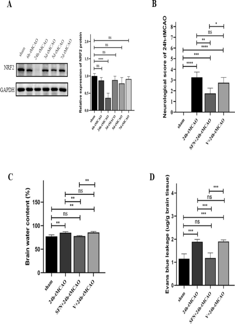
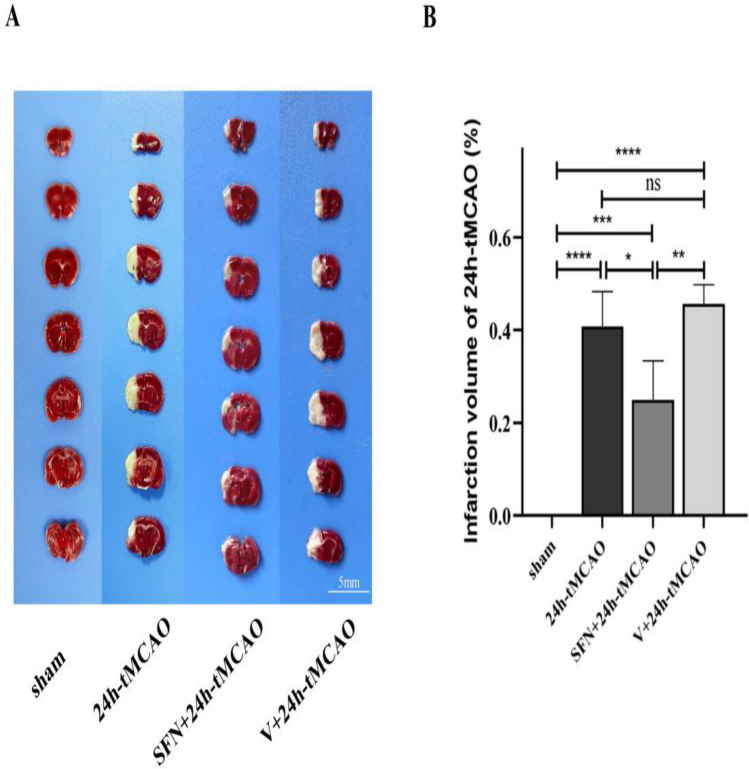
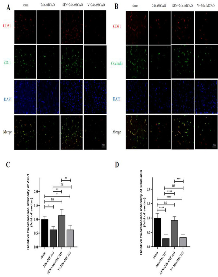
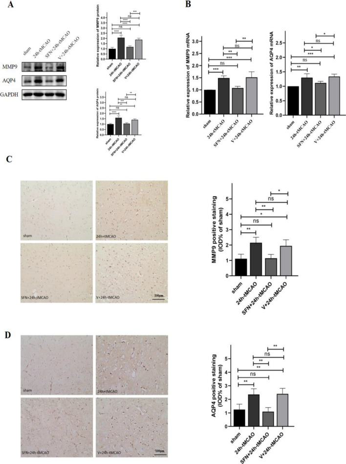
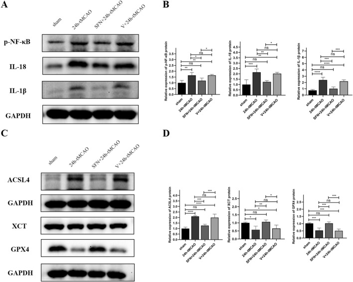
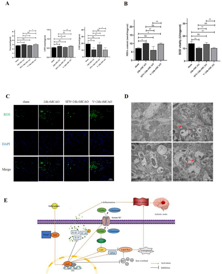
Arterial occlusion-induced ischemic stroke (IS) is a highly frequent stroke subtype. Nuclear factor erythroid 2-related factor 2 (NRF2) is a transcription factor that modulates antioxidant genes. Its role in IS is still unelucidated. The current study focused on constructing a transient middle cerebral artery occlusion (tMCAO) model for investigating the NRF2-related mechanism underlying cerebral ischemia/reperfusion (I/R) injury. Each male C57BL/6 mouse was injected with/with no specific NRF2 activator post-tMCAO. Changes in blood-brain barrier (BBB)-associated molecule levels were analyzed using western-blotting, PCR, immunohistochemistry, and immunofluorescence analysis. NRF2 levels within cerebral I/R model decreased at 24-h post-ischemia. NRF2 activation improved brain edema, infarct volume, and neurological deficits after MCAO/R. Similarly, sulforaphane (SFN) prevented the down-regulated tight junction proteins occludin and zonula occludens 1 (ZO-1) and reduced the up-regulated aquaporin 4 (AQP4) and matrix metalloproteinase 9 (MMP9) after tMCAO. Collectively, NRF2 exerted a critical effect on preserving BBB integrity modulating ferroptosis and inflammation. Because NRF2 is related to BBB injury regulation following cerebral I/R, this provides a potential therapeutic target and throws light on the underlying mechanism for clinically treating IS.

Keywords: Blood–brain barrier; Ferroptosis; Inflammation; Ischemic stroke; NRF2.


Conflict of interest statement

The authors declare no competing interests.

Figures

Figure 1
Figure 1
NRF2 expression in diverse time periods within mouse brain tissues post-tMCAO. NRF2 activation ameliorated cerebral ischemic reperfusion injury. (A) NRF2 protein expression detected by western blot (n = 4). (B) Neurological deficit scores in mice evaluated at 24 h post-tMCAO (n = 4). (C) Brain water content measured by the wet/dry approach for assessing brain edema (n = 4). (D) Evans blue dye leakage quantified using a spectrophotometer (n = 4). The results are indicated to be means ± SEM. *p < 0.05; **p < 0.01; ***p < 0.001; ****p < 0.0001 and N.S. nonsignificant versus the sham group.
Figure 2
Figure 2
NRF2 activator reduced infarction volume in mice brain tissue post-tMCAO. (A, B) Typical images showing TTC-stained brain sections and quantification of infarct volume in different groups (n = 4). Results are represented to be means ± SEM. *p < 0.05; **p < 0.01; ***p < 0.001; ****p < 0.0001 and N.S. nonsignificant versus the sham group. Scale bar = 5 mm.
Figure 3
Figure 3
NRF2 activation elevated ZO-1 and occludin levels within mouse brain tissue post-tMCAO. ZO-1 and occludin protein (A,B) and mRNA (C) expression of sham, 24 h-tMCAO, SFN + 24 h tMCAO, and V + 24 h- tMCAO groups (n = 4). (D) Immunohistochemical study of ZO-1 in the sham, 24 h-tMCAO, SFN + 24 h tMCAO, and V + 24 h tMCAO groups (n = 4). The results are suggested to be means ± SEM. *p < 0.05; **p < 0.01; ***p < 0.001; ****p < 0.0001 and N.S. nonsignificant versus the sham group. Scale bar = 100 µm.
Figure 4
Figure 4
SFN upregulates ZO-1 and occludin levels after tMCAO within mice. (A,B) Typical images showing double immunofluorescence staining with DAPI (blue)/ZO-1 (green) (A) and occludin (green) (B)/CD31 (red) colocalization (n = 4). (C,D) Immunofluorescence analysis of ZO1 and occludin (n = 4). The results indicate means ± SEM. *p < 0.05; **p < 0.01; ***p < 0.001; ****p < 0.0001 and N.S. nonsignificant versus the sham group. Scale bar = 100 µm.
Figure 5
Figure 5
NRF2 activation lowers MMP9 and AQP4 levels within mouse brain tissues post-tMCAO. MMP9 and AQP4 protein (A) and mRNA (B) expression of sham, 24 h-tMCAO, SFN + 24 h tMCAO, and V + 24 h tMCAO groups (n = 4). Immunohistochemical study of MMP9 (C) and AQP4 (D) of sham, 24 h-tMCAO, SFN + 24 h tMCAO, and V + 24 h tMCAO groups (n = 4). The results are indicated to be means ± SEM. *p < 0.05; **p < 0.01; ***p < 0.001; ****p < 0.0001 and N.S. nonsignificant versus the sham group. Scale bar = 100 µm.
Figure 6
Figure 6
SFN decreased MMP9 and AQP4 levels in mice post-tMCAO. (A,B) Typical images showing double immunofluorescence staining (n = 4) for DAPI (blue)/MMP9 (green) (A) and AQP4 (green) (B)/CD31 (red) colocalization. (C,D) Immunofluorescence analysis of MMP9 and AQP4 (n = 4). The results are suggested to be means ± SEM. *p < 0.05; **p < 0.01; ***p < 0.001; ****p < 0.0001 and N.S. nonsignificant versus the sham group. Scale bar = 100 µm.
Figure 7
Figure 7
SFN alleviated BBB disruption and brain damage by inhibiting inflammation and ferroptosis. (A,B) The p-NF-κB, IL-18, and IL-1β protein expression was measured with the application of WB (n = 4). (C,D) ACSL4, XCT, and GPX4 protein expression was measured with the application of WB (n = 4). Results are indicated to be means ± SEM. *p < 0.05; **p < 0.01; ***p < 0.001; ****p < 0.0001 and N.S. nonsignificant versus the sham group.
Figure 8
Figure 8
NRF2 activation suppressed ferroptosis in cerebral I/R mice. (A,B) Iron, LPO, GSH, MDA, and SOD levels were determined using relevant commercially available kits (n = 4). (C) Typical images showing ROS using the fluorescent probe DCFH-DA. Scale bar = 100 µm. (D) Typical TEM images in the peri-infarct region 24 h post-cerebral ischemic reperfusion. Red arrows suggest rupture of the neuronal outer mitochondrial membrane rapture and mitochondrial cristae reduction or disappearance. Scale bar = 1 µm. Results are suggested to be means ± SEM. *p < 0.05; **p < 0.01; ***p < 0.001; ****p < 0.0001 and N.S. nonsignificant versus the sham group. (E) Schematic of the study. Activation of NRF2 decreases cerebral I/R BBB damage by inhibiting ferroptosis and inflammation.

References

    1. Feigin VL, Stark BA, et al. Global, regional, and national burden of stroke and its risk factors, 1990–2019: A systematic analysis for the global burden of disease study 2019. Lancet Neurol. 2021;20:795–820. doi: 10.1016/S1474-4422(21)00252-0. - DOI - PMC - PubMed
    1. Saini V, Guada L, Yavagal DR. Global epidemiology of stroke and access to acute ischemic stroke interventions. Neurology. 2021;97:S6–S16. doi: 10.1212/WNL.0000000000012781. - DOI - PubMed
    1. Donkor ES. Stroke in the 21st century: A snapshot of the burden, epidemiology, and quality of life. Stroke Res. Treat. 2018;2018:3238165. doi: 10.1155/2018/3238165. - DOI - PMC - PubMed
    1. Daneman R, Engelhardt B. Brain barriers in health and disease. Neurobiol. Dis. 2017;107:1–3. doi: 10.1016/j.nbd.2017.05.008. - DOI - PubMed
    1. Ronaldson PT, Davis TP. Regulation of blood-brain barrier integrity by microglia in health and disease: A therapeutic opportunity. J. Cereb. Blood Flow Metab. 2020;40:S6–S24. doi: 10.1177/0271678X20951995. - DOI - PMC - PubMed

Substances