. 2024 Mar 4;14(1):5272.
doi: 10.1038/s41598-024-55983-w.

Elevation of astrocyte-derived extracellular vesicles over the first month post-stroke in humans

Affiliations

Elevation of astrocyte-derived extracellular vesicles over the first month post-stroke in humans

Matthew A Edwardson et al. Sci Rep. .

Abstract

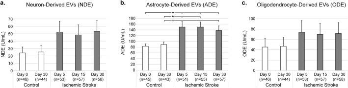
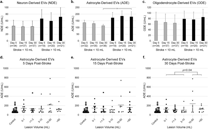
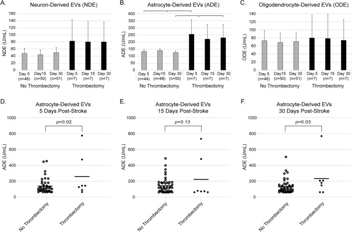
We sought to identify alterations in the quantity of plasma brain-derived extracellular vesicles (EV) over the first month post-stroke to shed light on related injury and repair mechanisms. We assessed plasma levels of presumed neuron-derived EVs (NDEs), astrocyte-derived EVs (ADEs), and oligodendrocyte-derived EVs (ODEs) in 58 patients 5, 15, and 30 days post-ischemic stroke and 46 controls matched for cardiovascular risk factors using sandwich immunoassays. Subsets of brain-derived EVs were identified by co-expression of the general EV marker CD9 and markers for neurons (L1CAM, CD171), astrocytes (EAAT1), and oligodendrocytes (MOG) respectively. Clinical MRIs assessed lesion volume and presence of hemorrhagic transformation. ADE levels were elevated 5, 15, and 30 days post-stroke compared to controls (p = 0.002, p = 0.002, and p = 0.005 respectively) with no significant change for NDE or ODE. ADEs were increased 15 days post-stroke in patients with hemorrhagic transformation (p = 0.04) compared to patients with no hemorrhage. We conclude that ADE levels are preferentially increased over the first month post-stroke in humans, possibly to provide trophic support to injured neurons following ischemia. ADEs hold potential as biomarkers of blood-brain barrier breakdown and hemorrhagic transformation, but this requires further study at earlier time points post-stroke.

Keywords: Astrocytes; Excitatory amino acid transporter 1; Exosomes; Extracellular vesicles; Hemorrhagic stroke; Ischemic stroke; Myelin oligodendrocyte glycoprotein; Neural cell adhesion molecule L1.


Conflict of interest statement

The study sponsor, NanoSomiX, Inc., provided support in the form of salaries for authors D.V. and M.M., and half the funding to isolate and quantify EV populations from plasma samples, but did not have any additional role in the study design, data collection and analysis, decision to publish, or preparation of the manuscript. NanoSomiX holds issued US and International pending patents on the methods for capture and analysis of plasma EVs used in this study. Georgetown University and NanoSomiX, Inc. have a collaborative agreement in place and have filed a provisional patent related to the use of brain-derived EVs in the care and diagnosis of stroke patients with M.A.E, M.M. and D.V. named as inventors. M.A.E. declares no other competing interests. This work was supported by internal funds from Georgetown University (M.A.E.) and support to analyze plasma samples was provided by NanoSomiX, Inc.

Figures

Figure 1
Figure 1
Extracellular vesicle (EV) concentrations (mean ± SE) in ischemic stroke versus control participants for (a) neuron-derived, (b) astrocyte-derived, and (c) oligodendrocyte-derived EVs (mean ± SE). There were no significant differences between the controls from day 0 to day 30. Wherever n < 46 for control or n < 58 for ischemic stroke there were samples that returned no measurable result. *p < 0.05; **p < 0.01.
Figure 2
Figure 2
Stroke volume < 10 mL versus > 10 mL in ischemic stroke participants and their corresponding extracellular vesicle (EV) concentrations (mean ± SE) 5, 15, and 30 days post-stroke for (a) neuron-derived, (b) astrocyte-derived, and (c) oligodendrocyte-derived EVs. Wherever n < 37 for volume < 10 mL or n < 21 for volume > 10 mL there were samples that returned no measurable result. Astrocyte-derived EV concentrations for individual participants (bar represents mean) at (d) 5 days, (e) 15 days, and (f) 30 days post-stroke. *p < 0.05.
Figure 3
Figure 3
Hemorrhagic transformation status for ischemic stroke patients and their corresponding extracellular vesicle (EV) concentrations (mean ± SE) at 5, 15, and 30 days post-stroke for (a) neuron-derived, (b) astrocyte-derived, and (c) oligodendrocyte-derived EVs. Wherever n < 46 for no hemorrhage there were samples that returned no measurable result. Astrocyte-derived EV (ADE) concentrations for individual participants (bar represents mean) at (d) 5 days, (e) 15 days, and (f) 30 days post-stroke. The 3 outliers with the highest ADE levels 15 days post-stroke are labeled in red, green, and blue to show how these outliers fluctuated over time. HI-1 hemorrhagic infarction type 1, HI-2 hemorrhagic infarction type 2, PH-1 parenchymal hematoma type 1 (listed in order of increasing severity). *p < 0.05.
Figure 4
Figure 4
Astrocyte-derived extracellular vesicle concentrations 5 days post-stroke versus ischemic stroke lesion volume, including designation of which participants had hemorrhagic transformation (triangles) and those with no hemorrhage (circles). Stroke lesion volume and hemorrhagic transformation were closely related, as evidenced by a chi-squared test with p = 0.002.
Figure 5
Figure 5
Mechanical thrombectomy status for ischemic stroke patients and their corresponding extracellular vesicle (EV) concentrations (mean ± SE) at 5, 15, and 30 days post-stroke for (a) neuron-derived, (b) astrocyte-derived, and (c) oligodendrocyte-derived EVs. Wherever n < 51 for no thrombectomy there were samples that returned no measurable result. Astrocyte-derived EV (ADE) concentrations for individual participants (bar represents mean) at (d) 5 days, (e) 15 days, and (f) 30 days post-stroke. *p < 0.05.

References

    1. Shah R, Patel T, Freedman JE. Circulating extracellular vesicles in human disease. N. Engl. J. Med. 2018;379:958–966. doi: 10.1056/NEJMra1704286. - DOI - PubMed
    1. Budnik V, Ruiz-Canada C, Wendler F. Extracellular vesicles round off communication in the nervous system. Nat. Rev. Neurosci. 2016;17:160–172. doi: 10.1038/nrn.2015.29. - DOI - PMC - PubMed
    1. Sun H, et al. Extracellular vesicles derived from astrocytes facilitated neurite elongation by activating the Hippo pathway. Exp. Cell Res. 2022;411:112937. doi: 10.1016/j.yexcr.2021.112937. - DOI - PubMed
    1. Zhuang X, et al. Treatment of brain inflammatory diseases by delivering exosome encapsulated anti-inflammatory drugs from the nasal region to the brain. Mol. Ther. 2011;19:1769–1779. doi: 10.1038/mt.2011.164. - DOI - PMC - PubMed
    1. Ohmichi T, et al. Quantification of brain-derived extracellular vesicles in plasma as a biomarker to diagnose Parkinson's and related diseases. Parkinson. Relat. Disord. 2019;61:82–87. doi: 10.1016/j.parkreldis.2018.11.021. - DOI - PubMed