. 2024 Mar 4;14(1):5371.
doi: 10.1038/s41598-024-55998-3.

Craniofacial bone anomalies related to cholesterol synthesis defects

Affiliations

Craniofacial bone anomalies related to cholesterol synthesis defects

Chihiro Iwaya et al. Sci Rep. .

Abstract

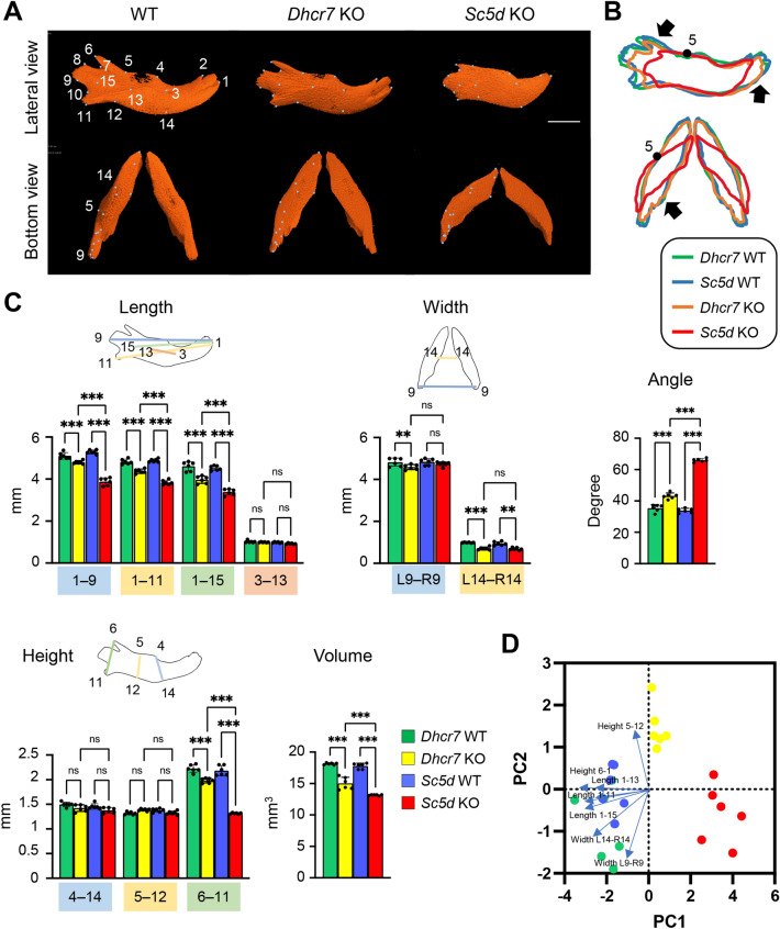
DHCR7 and SC5D are enzymes crucial for cholesterol biosynthesis, and mutations in their genes are associated with developmental disorders, which are characterized by craniofacial deformities. We have recently reported that a loss of either Dhcr7 or Sc5d results in a failure in osteoblast differentiation. However, it remains unclear to what extent a loss of function in either DHCR7 or SC5D affects craniofacial skeletal formation. Here, using micro computed tomography (μCT), we found that the bone phenotype differs in Dhcr7-/- and Sc5d-/- mice in a location-specific fashion. For instance, in Sc5d-/- mice, although craniofacial bones were overall affected, some bone segments, such as the anterior part of the premaxilla, the anterior-posterior length of the frontal bone, and the main body of the mandible, did not present significant differences compared to WT controls. By contrast, in Dhcr7-/- mice, while craniofacial bones were not much affected, the frontal bone was larger in width and volume, and the maxilla and palatine bone were hypoplastic, compared to WT controls. Interestingly the mandible in Dhcr7-/- mice was mainly affected at the condylar region, not the body. Thus, these results help us understand which bones and how greatly they are affected by cholesterol metabolism aberrations in Dhcr7-/- and Sc5d-/- mice.


Conflict of interest statement

The authors declare no competing interests.

Figures

Figure 1
Figure 1
µCT analysis of the premaxilla. (A) 3D reconstruction of the premaxilla in E18.5 WT, Sc5d KO, and Dhcr7 KO mice. Definitions of landmarks: 1. most anterior superior point of the premaxilla; 2. most lateral point of the premaxillary-maxillary suture; 3. tip of the frontal process of the premaxilla; 4. most medial point of the premaxillary-maxillary suture; 5. most anterior point of the anterior palatine foramen; 6. most posterior point of the premaxilla; and 7. most posterior point of the incisive foramen. Scale bar: 1 mm. (B) Wiring trace of the premaxilla in E18.5 WT (blue), Dhcr7 KO (orange), and Sc5d KO (red) mice. Arrows indicate the missing portion in Sc5d KO mice. (C) Quantification of the size (length, width, height, and volume) of the maxilla from Dhcr7 WT (green bars), Dhcr7 KO (yellow bar), Sc5d WT (blue bars), and Sc5d KO (red bars) mice. *p < 0.05; **p < 0.01; ***p < 0.001; ns, not significant. N/A, not available. (D) Scatter plots of individual scores of PCA displaying the degree of morphological variances (length, width, height, and volume) of the premaxilla in Dhcr7 WT (green dots), Dhcr7 KO (yellow dots), Sc5d WT (blue dots), and Sc5d KO (red dots) mice, shown by PC1 and PC2. Distribution in mutants (Sc5d KO and Dhcr7 KO) and control littermates (Sc5d WT and Dhcr7 WT) along with 10 principal components (blue arrows) are shown.
Figure 2
Figure 2
µCT analysis of the maxilla. (A) 3D reconstruction of the maxilla in E18.5 WT, Dhcr7 KO, and Sc5d KO mice. Definitions of landmarks: 1. anterior point of the maxilla; 2. lateral inferior intersection of the frontal and zygomatic process of the maxilla; 3. tip of the zygomatic process of the maxilla; 4. anterior-medial point to the zygomatic process of the maxilla; 5. posterior point of the maxilla; 6. posterior-lateral point of the palatine process of the maxilla; 7. posterior-medial point of the palatine process of the maxilla; 8. most anterior-medial point of the palatine process of the maxilla; 9. anterior-lateral point of the palatine process of the maxilla; and 10. medial point of the premaxillary-maxillary suture. Scale bar: 1 mm. (B) Wiring trace of the maxilla in E18.5 WT (blue), Dhcr7 KO (orange), and Sc5d KO (red) mice. Arrows indicate the missing portion in Sc5d KO mice. (C) Quantification of the size (length, width, height, right-left distance, and volume) of the maxilla from Dhcr7 WT (green bars), Dhcr7 KO (yellow bar), Sc5d WT (blue bars), and Sc5d KO (red bars) mice. *p < 0.05; **p < 0.01; ***p < 0.001; ns, not significant. N/A, not available. (D) Scatter plots of individual scores of PCA displaying the degree of morphological variances (length, width, height, right-left distance, and volume) of the maxilla in Dhcr7 WT (green dots), Dhcr7 KO (yellow dots), Sc5d WT (blue dots), and Sc5d KO (red dots) mice, shown by PC1 and PC2. Distributions in mutant (Sc5d KO and Dhcr7 KO) and control littermate (Sc5d WT and Dhcr7 WT) mice along with 14 principal components (blue arrows) are shown.
Figure 3
Figure 3
µCT analysis of the palatine bone. (A) 3D reconstruction of the palatine bone in E18.5 WT, Dhcr7 KO, andSc5d KO mice. Definitions of landmarks: 1. most anterior-lateral point of the palatine plate; 2. tip of the orbital process of the palatine bone; 3. lateral point of the palatine bone; 4. posterior point of the palatine bone; 5. posterior-medial point of the horizontal plate of the palatine bone; 6. anterior-medial point of the horizontal plate of the palatine bone; and 7. anterior superior point of the perpendicular plate. Scale bar: 1 mm. (B) Wiring trace of the palatine bone in E18.5 WT (blue), Dhcr7 KO (orange), and Sc5d KO (red) mice. Arrows indicate the missing portion in Sc5d KO mice. (C) Quantification of the size (length, width, height, right-left distance, and volume) of the palatine bone from Dhcr7 WT (green bars), Dhcr7 KO (yellow bar), Sc5d WT (blue bars), and Sc5d KO (red bars) mice. *p < 0.05; **p < 0.01; ***p < 0.001; ns, not significant. (D) Scatter plots of individual scores of PCA displaying the degree of morphological variances (length, width, height, right-left distance and volume) of the palatine in Dhcr7 WT (green dots), Dhcr7 KO (yellow dots), Sc5d WT (blue dots), and Sc5d KO (red dots) mice, shown by PC1 and PC2. Distribution in mutants (Sc5d KO and Dhcr7 KO) and control littermates (Sc5d WT and Dhcr7 WT) along with 10 principal components (blue arrows) are shown.
Figure 4
Figure 4
µCT analysis of the frontal bone. (A) 3D reconstruction of the frontal bone in E18.5 WT, Dhcr7 KO, and Sc5d KO mice. Definitions of landmarks: 1. most anterior–superior point of the frontal bone; 2. most posterior-superior point of the frontal bone; 3. most posterior-lateral intersection of the frontal bone and parietal bone; 4. most posterior-inferior point of the frontal bone; 5. most anterior-inferior point of the frontal bone; and 6. midpoint of the interfrontal suture. Scale bar: 1 mm. (B) Wiring trace of the frontal bone in E18.5 WT (blue), Dhcr7 KO (orange), and Sc5d KO (red) mice. Arrows indicate the missing portion in Sc5d KO mice. (C) Quantification of the size (length, width, height, right-left distance, and volume) of the frontal bone from Dhcr7 WT (green bars), Dhcr7 KO (yellow bar), Sc5d WT (blue bars), and Sc5d KO (red bars) mice. *p < 0.05; **p < 0.01; ***p < 0.001; ns, not significant. (D) Scatter plots of individual scores of PCA displaying the degree of morphological variances (length, width, height, right-left distance and volume) of the frontal bone in Dhcr7 WT (green dots), Dhcr7 KO (yellow dots), Sc5d WT (blue dots), and Sc5d KO (red dots) mice, shown by PC1 and PC2. Distributions in mutant (Sc5d KO and Dhcr7 KO) and control littermate (Sc5d WT and Dhcr7 WT) mice along with 9 principal components (blue arrows) are shown.
Figure 5
Figure 5
µCT analysis of the mandible. (A) 3D reconstruction of the mandible in E18.5 WT, Dhcr7 KO, and Sc5d KO mice. Definitions of landmarks: 1. most anterior point of the mandible; 2. anterior–superior point of the mandible; 3. mental foramen; 4. molar alveolus of dentary; 5. anterior junction of the mandibular ramus and body; 6. superior tip of the coronary process of the mandible; 7. most inferior point of the mandibular notch; 8. anterior point of the condylar process of the mandible; 9. posterior point of the condylar process of the mandible; 10. superior point of the angular process of the mandible; 11. secondary cartilage of the angular process of the mandible; 12. inferior junction of the mandibular ramus and body; 13. midpoint of the external oblique ridge; 14. inferior point of the mandibular body; and 15. mandibular foramen. Scale bar: 1 mm. (B) Wiring trace of the mandible in E18.5 WT (blue), Dhcr7 KO (orange), and Sc5d KO (red) mice. Arrows indicate the missing portion in Sc5d KO mice. (C) Quantification of the size (length, width, height, angle, and volume) of the mandible from Dhcr7 WT (green bars), Dhcr7 KO (yellow bar), Sc5d WT (blue bars), and Sc5d KO (red bars) mice. *p < 0.05; **p < 0.01; ***p < 0.001; ns, not significant. (D) Scatter plots of individual scores of PCA displaying the degree of morphological variances (length, width, height, angle, and volume) of the mandible in Dhcr7 WT (green dots), Dhcr7 KO (yellow dots), Sc5d WT (blue dots), and Sc5d KO (red dots) mice, shown by PC1 and PC2. Distribution in mutants (Sc5d KO and Dhcr7 KO) and control littermates (Sc5d WT and Dhcr7 WT) along with 11 principal components (blue arrows) are shown.

Similar articles

Cited by

References

    1. Porter FD, Herman GE. Malformation syndromes caused by disorders of cholesterol synthesis. J. Lipid. Res. 2011;52:6–34. doi: 10.1194/jlr.R009548. - DOI - PMC - PubMed
    1. Dietschy JM, Turley SD, Spady DK. Role of liver in the maintenance of cholesterol and low density lipoprotein homeostasis in different animal species, including humans. J. Lipid. Res. 1993;34:1637–1659. doi: 10.1016/S0022-2275(20)35728-X. - DOI - PubMed
    1. Suzuki A, Minamide M, Iwaya C, Ogata K, Iwata J. Role of metabolism in bone development and homeostasis. Int. J. Mol. Sci. 2020 doi: 10.3390/ijms21238992. - DOI - PMC - PubMed
    1. Krakowiak PA, et al. Lathosterolosis: An inborn error of human and murine cholesterol synthesis due to lathosterol 5-desaturase deficiency. Hum. Mol. Genet. 2003;12:1631–1641. doi: 10.1093/hmg/ddg172. - DOI - PubMed
    1. Porter FD. Smith-Lemli-Opitz syndrome: Pathogenesis, diagnosis and management. Eur. J. Hum. Genet. 2008;16:535–541. doi: 10.1038/ejhg.2008.10. - DOI - PubMed