. 2024 Mar 4;7(1):264.
doi: 10.1038/s42003-024-05934-y.

Topical application of calcitonin gene-related peptide as a regenerative, antifibrotic, and immunomodulatory therapy for corneal injury

Affiliations

Topical application of calcitonin gene-related peptide as a regenerative, antifibrotic, and immunomodulatory therapy for corneal injury

Asmaa A Zidan et al. Commun Biol. .

Abstract

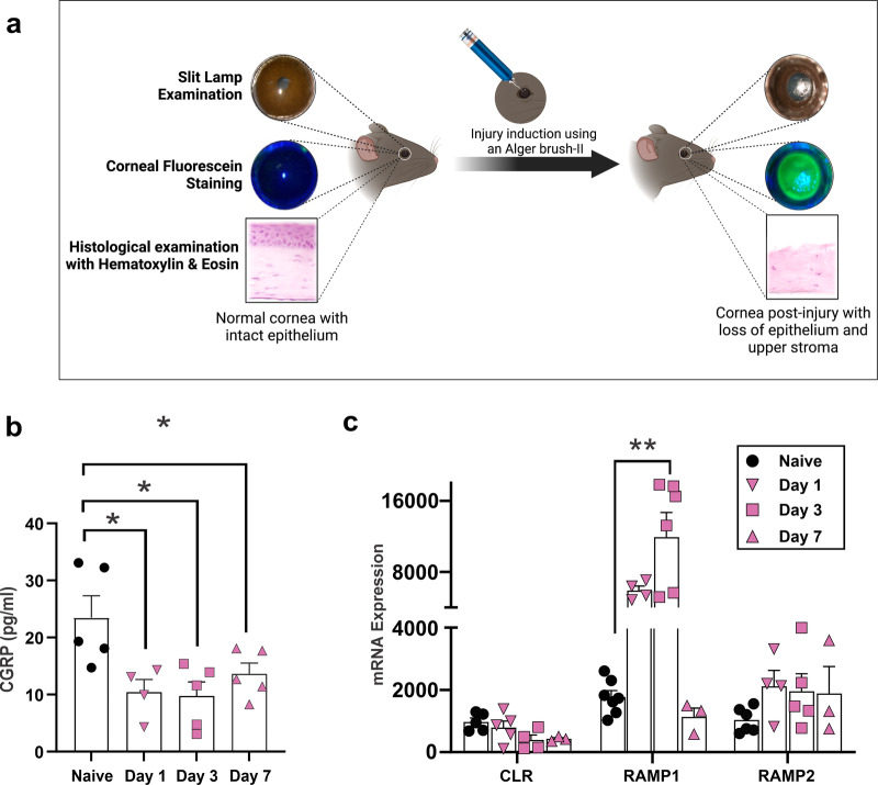
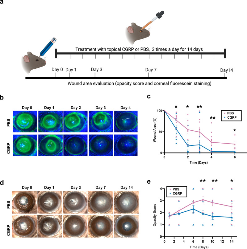
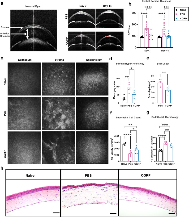
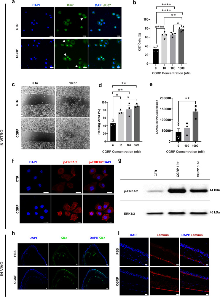
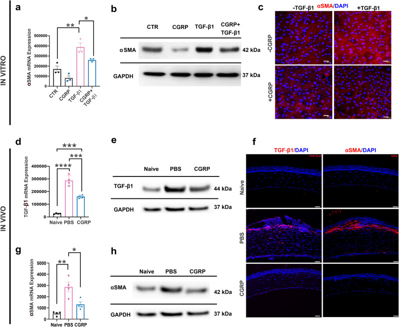
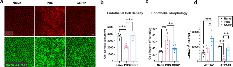
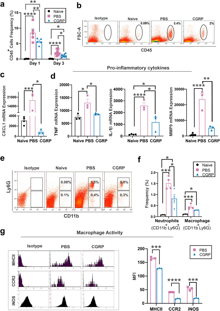
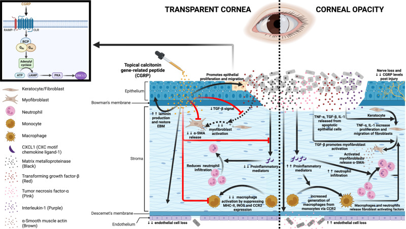
Calcitonin gene-related peptide (CGRP) is a multifunctional neuropeptide abundantly expressed by corneal nerves. Using a murine model of corneal mechanical injury, we found CGRP levels in the cornea significantly reduced after injury. Topical application of CGRP as an eye drop accelerates corneal epithelial wound closure, reduces corneal opacification, and prevents corneal edema after injury in vivo. CGRP promotes corneal epithelial cell migration, proliferation, and the secretion of laminin. It reduces TGF-β1 signaling and prevents TGF-β1-mediated stromal fibroblast activation and tissue fibrosis. CGRP preserves corneal endothelial cell density, morphology, and pump function, thus reducing corneal edema. Lastly, CGRP reduces neutrophil infiltration, macrophage maturation, and the production of inflammatory cytokines in the cornea. Taken together, our results show that corneal nerve-derived CGRP plays a cytoprotective, pro-regenerative, anti-fibrotic, and anti-inflammatory role in corneal wound healing. In addition, our results highlight the critical role of sensory nerves in ocular surface homeostasis and injury repair.


Conflict of interest statement

The authors declare no competing interests.

Figures

Fig. 1
Fig. 1. Mechanical injury causes a decrease in CGRP levels in the cornea.
a Schematic figure showing the method of mechanical injury. The epithelium and superficial stroma were removed using Algerbrush-II. b Assessment of CGRP protein levels using ELISA showed significantly reduced expression following corneal injury on days 1, 3, and 7. c The gene expression levels of the CGRP receptors, calcitonin receptor-like receptor (CLR), receptor activity-modifying protein (RAMP) 1, and RAMP2. RT-PCR showed upregulation of the RAMP1 on day 3 post-injury and returned to normal levels by day 7. (n = 3–5 per group). The data were presented as mean ± standard error of mean (SEM) and comparison is determined by a one-way ANOVA test with pairwise comparison. *p < 0.05, **p < 0.01. (Legend: Circle = naive, Inverted Triangle = Day 1, Square = Day 3, Upright Triangle = Day 7). Figure 1a was created with BioRender.com.
Fig. 2
Fig. 2. Topically applied CGRP accelerates corneal epithelial wound closure and reduces corneal opacity.
a Schematic figure showing the timeline of treatment and clinical examination following injury. b Corneal fluorescein staining was performed to compare the size of epithelial defects in CGRP- and PBS-treated mice at different time points post-injury. c The assessment of the wound area shows that CGRP treatment resulted in a significantly smaller wound area compared to PBS-treated controls across all time points from 24 h to 6 days (n = 9 per group). d Representative slit lamp photographs of PBS and CGRP-treated eyes up to 14 days after injury. The corneas of the PBS-treated controls showed progressive stromal opacification, whereas CGRP treatment showed significantly lower corneal opacity. e The scoring of corneal opacity was performed in a blinded fashion and showed a significantly lower score in CGRP-treated mice (n = 12 per group). The data were represented as mean ± SEM. The statistical significance was determined by unpaired t-test, *p < 0.05, **p < 0.01. (Legend: Pink Square = PBS, Blue Triangle = CGRP). Figure 2a was created with BioRender.com.
Fig. 3
Fig. 3. CGRP decreases corneal thickness, scar formation, and endothelial cell loss after injury.
a Representative AS-OCT images showed a significant increase in central corneal thickness (CCT) and stromal hyperreflectivity in PBS-treated mice, whereas they were comparable to naïve mice in the treated group on days 7 and 14 post-injury. b CGRP treatment resulted in significantly lower CCT compared to PBS-treated controls on days 7 and 14 (n = 10 per group). c Representative IVCM images of the corneal epithelium, stroma, and endothelium. The analysis of IVCM images showed that CGRP treatment resulted in decreased stromal hyperreflectivity (d), scar depth (e), endothelial cell loss (f), and endothelial coefficient of variation (g) compared to PBS-treated mice at day 14 post-injury. h The histological analysis by hematoxylin and eosin staining showed reduced corneal thickness and inflammatory cell infiltration following CGRP compared to the PBS treatment (scale bar = 200 µm). d n = 5, eg n = 6 per group). The data were represented as mean ± SEM, and the statistical significance was determined by one-way ANOVA (b, d, f, g) and unpaired t-test (e), *p < 0.05, **p < 0.01, ***P < 0.001, ****P < 0.0001. (Legend: Black Circle = Naïve, Pink Square = PBS, Blue Triangle = CGRP).
Fig. 4
Fig. 4. CGRP promotes corneal epithelial cell (CEC) regeneration in vitro and in vivo.
a Human CEC were cultured and CGRP (1 μM for 24 h) led to an increased frequency of proliferating Ki67+ cells (arrowheads, green) (scale bar = 20 µm). b CGRP resulted in an increase in the Ki67+ cells  in a dose-dependent manner (n = 4 per group). c CEC were cultured to a monolayer and a linear scratch was created. d CGRP promoted CEC migration in a dose-dependent manner (n = 3 per group). e RT-PCR data showed significantly increased laminin 332expression in CEC by CGRP (n = 3 per group). f CGRP (1 μM for 1 h) increased the phosphorylation of ERK (p-ERK antibody, red, (scale = 20 µm). g Western blot analysis confirmed the increased p-ERK levels with CGRP treatment. h Mouse corneas obtained (on day 4) from the CGRP-treated mice showed more Ki67 (green) staining in the epithelium compared to PBS-treated controls, (scale bar = 100 µm). i A higher level of laminin immunostaining (red) was also observed in the cornea derived from CGRP-treated mice compared to the PBS-treated controls (scale bar = 20 µm). The data were presented as mean ± SEM, and the statistical significance was determined by one-way ANOVA with pairwise comparison, *p < 0.05, **p < 0.01, ****P < 0.0001.
Fig. 5
Fig. 5. CGRP suppresses TGF-β1 signaling and corneal stromal fibroblast activation in vitro and in vivo.
Murine corneal fibroblasts were cultured in a medium supplemented with 10 ng/ml TGF-β1 for 24 h, in the presence or absence of 1 µM CGRP. CGRP significantly decreased the TGF-β1-mediated expression of α-SMA assessed via RT-PCR (a), western blot (b), and immunostaining (c) in vitro (n = 3 per group). Corneas derived from CGRP-treated mice showed significantly lower expression of TGF-β1 compared to PBS-treated controls in vivo in RT-PCR (d, n = 4 per group), western blot (e), and immunostaining (f, scale = 50 μm). Similarly, α-SMA levels in RT-PCR (g, n = 4 per group), western blot (h), and immunostaining (f) were significantly elevated post-injury and decreased by CGRP treatment in vivo. The data were presented as mean ± SEM, and the statistical significance was determined by one-way ANOVA with pairwise comparison. *p < 0.05, **p < 0.01, ***P < 0.001, ****P < 0.0001. (Legend in d, g: Black Circle = Naïve, Pink Square = PBS, Blue Triangle = CGRP).
Fig. 6
Fig. 6. CGRP preserves corneal endothelial cell density and function in vivo.
a Mouse corneas were collected on Day 14 post-injury and CGRP treatment led to preserved zonula occludens-1 (ZO-1) and Na+/K+ ATPase staining, compared to the PBS-treated controls (scale = 20 μm). Analysis of immunohistochemical images showed higher endothelial cell density (b) and lower coefficient of variation (c) in vivo (n = 6 per group). d RT-PCR evaluation of mouse corneas showed higher gene expression of α1 and α3 isoforms of Na+/K+ ATPase in CGRP-treated mice compared to controls (n = 3 per group). The data were presented as mean ± SEM, and the statistical significance was determined by one-way ANOVA with pairwise comparison (bd), *p < 0.05, **p < 0.01, ***P < 0.001. (Legend: Black Circle = Naïve, Pink Square = PBS, Blue Triangle = CGRP).
Fig. 7
Fig. 7. CGRP dampens tissue inflammation after injury in vivo.
a Lower frequencies of CD45+ cells in corneas derived from CGRP-treated mice compared to controls on day 1 and day 3 post-injury, (n = 5 per group). b Representative flow cytometry plots show that CGRP treatment suppressed the infiltration of CD45+ into the cornea on day 3 post-injury compared to PBS-treated controls. The RT-PCR analysis showed that the CGRP treatment resulted in significantly lower expression of CXCL1 (c), IL-1β, TNF, and MMP-9 (d) on day 3 post-injury (n = 3 per group). e Representative flow cytometry plot shows the frequencies of neutrophils (CD11b+Ly6G+) and macrophage (CD11b+Ly6G-) in the mice cornea. f The frequencies of neutrophils were significantly lower in corneas derived from CGRP-treated mice compared to controls, and the frequencies of macrophages were comparable in the two groups (n = 3 per naive group, n = 4 per PBS and CGRP group). g CGRP treatment resulted in significant suppression of MCH-II, CCR2, and iNOS expression in CGRP-treated mice compared to the controls (n = 3 per group). The data were presented as mean ± SEM, and the statistical significance was determined by one-way ANOVA with pairwise comparison (af) and unpaired t-test (g), *p < 0.05, **p < 0.01, ***P < 0.00, ****P < 0.0001. (Legend: Black Circle = Naïve, Pink Square = PBS, Blue Triangle = CGRP).
Fig. 8
Fig. 8. Schematic showing the effects of CGRP on corneal wound healing after mechanical injury.
Injury leads to nerve damage and a decrease in CGRP level in the cornea. Topical application of CGRP promotes corneal epithelial cell regeneration and restores the epithelial basement membrane, thus reducing the release of pro-inflammatory and pro-fibrotic mediators including TNF-α, TGF-β, IL-1, and CXCL1 into the stroma. This leads to reduced keratocyte activation and stromal fibrosis. In addition, CGRP reduces neutrophil infiltration, macrophage maturation, and the production of inflammatory cytokines. It reduces corneal endothelial cell loss and maintains its pump function. Clinically, topical application of CGRP as an eye drop accelerates epithelial closure, preserves transparency, and prevents scar formation and edema after corneal injury. The figure was created with BioRender.com.

Update of

Similar articles

Cited by

References

    1. Wang, E. et al. Global trends in blindness and vision impairment due to corneal opacity 1984-2020: a meta-analysis. Ophthalmology10.1016/j.ophtha.2023.03.012 (2023). - PMC - PubMed
    1. Jeng BH, Ahmad S. In pursuit of the elimination of corneal blindness: is establishing eye banks and training surgeons enough? Ophthalmology. 2021;128:813–815. doi: 10.1016/j.ophtha.2020.06.042. - DOI - PubMed
    1. McGhee CN, Dean S, Danesh-Meyer H. Locally administered ocular corticosteroids: benefits and risks. Drug Safety. 2002;25:33–55. doi: 10.2165/00002018-200225010-00004. - DOI - PubMed
    1. Gain P, et al. Global survey of corneal transplantation and eye banking. JAMA Ophthalmol. 2016;134:167–173,. doi: 10.1001/jamaophthalmol.2015.4776. - DOI - PubMed
    1. Tan DT, Dart JK, Holland EJ, Kinoshita S. Corneal transplantation. Lancet. 2012;379:1749–1761. doi: 10.1016/S0140-6736(12)60437-1. - DOI - PubMed

Substances