. 2024 Jun;45(6):1160-1174.
doi: 10.1038/s41401-024-01234-7. Epub 2024 Mar 4.

α4 nicotinic receptors on GABAergic neurons mediate a cholinergic analgesic circuit in the substantia nigra pars reticulata

Affiliations

α4 nicotinic receptors on GABAergic neurons mediate a cholinergic analgesic circuit in the substantia nigra pars reticulata

Yu Han et al. Acta Pharmacol Sin. 2024 Jun.

Abstract

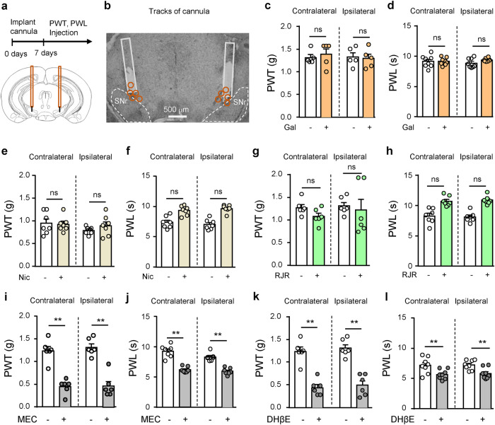
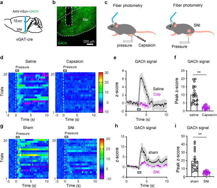
Nicotinic acetylcholine receptors (nAChRs) regulate pain pathways with various outcomes depending on receptor subtypes, neuron types, and locations. But it remains unknown whether α4β2 nAChRs abundantly expressed in the substantia nigra pars reticulata (SNr) have potential to mitigate hyperalgesia in pain states. We observed that injection of nAChR antagonists into the SNr reduced pain thresholds in naïve mice, whereas injection of nAChR agonists into the SNr relieved hyperalgesia in mice, subjected to capsaicin injection into the lower hind leg, spinal nerve injury, chronic constriction injury, or chronic nicotine exposure. The analgesic effects of nAChR agonists were mimicked by optogenetic stimulation of cholinergic inputs from the pedunculopontine nucleus (PPN) to the SNr, but attenuated upon downregulation of α4 nAChRs on SNr GABAergic neurons and injection of dihydro-β-erythroidine into the SNr. Chronic nicotine-induced hyperalgesia depended on α4 nAChRs in SNr GABAergic neurons and was associated with the reduction of ACh release in the SNr. Either activation of α4 nAChRs in the SNr or optogenetic stimulation of the PPN-SNr cholinergic projection mitigated chronic nicotine-induced hyperalgesia. Interestingly, mechanical stimulation-induced ACh release was significantly attenuated in mice subjected to either capsaicin injection into the lower hind leg or SNI. These results suggest that α4 nAChRs on GABAergic neurons mediate a cholinergic analgesic circuit in the SNr, and these receptors may be effective therapeutic targets to relieve hyperalgesia in acute and chronic pain, and chronic nicotine exposure.

Keywords: cholinergic system; chronic nicotine; nicotinic acetylcholine receptor; pain; pedunculopontine nucleus; substantia nigra pars reticulata.


Conflict of interest statement

The authors declare no competing interests.

Figures

Fig. 1
Fig. 1. Inhibition of nAChRs in the SNr reduces pain threshold in mice.
a, b Diagram and a representative image showing injection of drugs into the substantia nigra pars reticulata (SNr) through cannulas. Orange circles represent locations of cannulas. c, d Mechanical paw withdrawal threshold (PWT) and thermal paw withdrawal latency (PWL) on both hind paws after saline (-) (n = 7) or galatamine (+) (Gal, 10 μM, 300 nl) (n = 7) injection into the SNr. e, f PWT and PWL on both hind paws after saline (-) (n = 7) or nicotine (+) (Nic, 1 μM, 300 nl) (n = 7) injection into the SNr. g, h PWT and PWL on both hind paws after saline (-) (n = 7) or RJR-2403 (+) (RJR, 500 μM, 300 nl) (n = 7) injection into the SNr. i, j PWT and PWL on both hind paws after saline (-) (n = 7) or mecamylamine (+) (MEC, 100 μM, 300 nl) (n = 7) injection into the SNr. k, l PWT and PWL on both hind paws after saline (-) (n = 7) or DHβE (+) (0.3 μM, 300 nl) (n = 7) injection into the SNr. Two-tailed paired t-tests were used for (cl). n = 7. ‘ns’ not significant. **P < 0.01.
Fig. 2
Fig. 2. Nicotine in the SNr mitigates hyperalgesia in acute and chronic pain mouse models.
a Time line and schematic diagram of drug injection into the substantia nigra pars reticulata (SNr) through a cannula. b Representative images showing injection of Alexa 488-conjugated Ctb into the SNr. Orange circles represent locations of cannulas. c Pain mouse models: chronic constriction injury (CCI) (n = 7) and spared nerve injury (SNI) (n = 7) of the sciatic nerve. d, e PWT and PWL on both hind paws of naïve and Cap mice 15–30 and 45–60 min after Cap injection and 15 min after injection of saline (Nic -) or nicotine (Nic +, 1 μM, 300 nl) into the SNr. f Time courses of PWT and PWL in sham mice (n = 7) and CCI mice (n = 7) before and after surgery. g PWT and PWL in CCI mice before and two weeks after surgery (without or with Nic injection into the SNr). h Time courses of PWT and PWL in sham mice (n = 7) and SNI mice (n = 7) before and after surgery. i PWT and PWL in SNI mice before and two weeks after surgery (without or with Nic injection into the SNr). One-way repeated measures ANOVAs with Tukey’s multiple comparison tests were used in (d, e, g, i). Two-way repeated measures ANOVAs were used in (f, h). ** P < 0.01.
Fig. 3
Fig. 3. Activation of α4β2 nAChRs mitigates hyperalgesia in acute and chronic pain.
a Diagram of drug injection into the SNr. b Representative images showing c-Fos-(+) SNr neurons in mice subjected to saline and RJR-2403 (RJR, 500 μM) injection. c Summary of c-Fos-(+) SNr neurons on both sides after saline or RJR injection. Contralateral, t = 6.37, P < 0.0001, n = 6; Ipsilateral, t = 13.25, P < 0.0001, n = 6. Two-tailed t tests. d, e PWT and PWL on both hind paws of Cap mice 15–30 and 45–60 min after Cap injection and 30 min after injection of saline(-) or RJR(+) into the SNr. f, g PWT and PWL on both hind paws of Cap mice (15–30 and 45–60 min after Cap injection) after injection of saline (-) or RJR (+) or RJR + DHβE into the SNr. h, i PWT and PWL after injection of saline (-) or RJR (+) into the SNr of CCI mice. j, k PWT and PWL after injection of saline (-) or RJR (+) into the SNr of SNI mice. l, m PWT and PWL after injection of saline (-) or ropinirole (Rop) or RJR + Rop into the SNr of CCI mice. One-way repeated measures ANOVAs with Bonferroni’s multiple comparison tests were used in (dm). ‘ns’ not significant. * P < 0.05. ** P < 0.01.
Fig. 4
Fig. 4. α4 nAChRs in SNr GABAergic neurons modulate pain states in mice.
a Diagram for downregulation of α4 nAChRs in SNr GABAergic neurons. b, c The specificity (left histogram) and efficiency (right histogram) of viral labeling of SNr GABAergic neurons. d mRNA of Vgat, α4, β2, α7, and Th in eGFP-labeled SNr GABAergic neurons in shRNA-injected mice (n = 6) and scrRNA-injected mice (n = 6). e, f 50 μM RJR-2403 (RJR)-evoked inward currents in SNr GABAergic neurons and SNc non-GABAergic neurons from shRNA (n = 5) and naive mice (n = 5). g PWT and PWL measured (15–30 min) before and after RJR injection into the SNr in shRNA mice (n = 7) subjected to Cap injection on the lower leg. h PWT and PWL measured (45–60 min) before and after RJR injection into the SNr in shRNA mice (n = 7) subjected to Cap injection on the lower leg. i PWT and PWL measured (15–30 min) before and after Nic injection into the SNr in shRNA mice (n = 7) subjected to Cap injection on the lower leg. j PWT and PWL measured (45–60 min) before and after RJR injection into the SNr in shRNA mice (n = 7) subjected to Cap injection on the lower leg. k PWT and PWL before and after injection of RJR into the SNr in shRNA (n = 6) mice subjected to SNI. l PWT and PWL before and after injection of Nic into the SNr in shRNA mice (n = 6) subjected to SNI. Two-tailed t tests were used in (d, f). One-way repeated measures ANOVAs with Tukey’s multiple comparisons tests were used in (gl). ‘ns’ not significant. * P < 0.05. ** P < 0.01.
Fig. 5
Fig. 5. Stimulating the PPN-SNr projection mitigates hyperalgesia in acute pain.
a Diagram and representative images showing optogenetic stimulation of the cholinergic projection from the pedunculopontine nucleus (PPN) to the SNr. b Summary of ChR2- and ChAT antibody-labeled PPN neurons. c A representative image showing an optical fiber in the SNr. d, e PWT and PWL on both hind paws before, during, and after blue light illumination of the PPN-SNr projection in ChR2 mice (n = 15). f, g PWT and PWL on both hind paws before, during, and after blue light illumination of the SNr in eYFP mice (n = 7). h, i PWT and PWL on both hand paws in Cap mice (n = 8) (45–60 min after Cap injection) before and during optogenetic stimulation of the PPN-SNr projection. j, k PWT and PWL on both hand paws in eYFP mice (n = 7) (45–60 min after Cap injection) before and during blue light illumination of the SNr. l, m PWT and PWL in Cap mice before and during optogenetic stimulation of the PPN-SNr projection without or with i.p. injection of mecamylamine (MEC) (n = 6). n, o PWT and PWL in Cap mice before and during optogenetic stimulation of the PPN-SNr projection without or with i.p. injection of DHβE (n = 6). p, q PWT or PWL of SNI mice before and during optogenetic stimulation of the PPN-SNr projection (n = 8). One-way ANOVAs with Tukey’s multiple comparison tests were used in (dq). ‘ns’ not significant. ** P < 0.01.
Fig. 6
Fig. 6. The role of α4 nAChRs on SNr GABAergic neurons in chronic nicotine-induced hyperalgesia in mice.
a Diagram for chronic subcutaneous injection of nicotine (Nic) or saline in mice and PWT and PWL was measured 5 h after injection. b, c PWT and PWL in chronic Nic mice (n = 8) and saline mice (n = 8). d, e AAV-CMV-DIO-Chrna4-eGFP was transfected in SNr GABAergic neurons. f mRNA levels of Vgatα4, β2, α7, and Th in eGFP-labeled SNr GABAergic neurons in Chrna4 mice and eGFP mice was measured with quantitative real-time polymerase chain reaction (qRT-PCR). g, h Representative traces and summary of RJR-evoked inward currents in SNr GABAergic and SNc non-GABAergic neurons from Chrna4 and eGFP mice. i Comparison of PWT and PWL in eGFP, Chrna4, and shRNA mice. j PWT and PWL in shRNA mice (n = 7) and scrRNA mice (n = 15) subjected to daily subcutaneous injection of Nic. k Diagram of chronic nicotine treatment, injection of Nic or RJR into the SNr, and pain behavior tests. l, m PWT in chronic nicotine mice before and after injection of Nic (n = 8) or RJR (n = 8) into the SNr. Two-way repeated measures ANOVAs were used in (b, c, j). Two-tailed t tests were used in (f, h). One-way ANOVA was used in (i). One-way repeated measures ANOVAs with Tukey’s multiple comparison tests were used in (i, l, m).
Fig. 7
Fig. 7. Chronic nicotine reduces ACh release in the SNr.
a, b Diagram and a representative image for fiber photometry recording of fluorescence intensity of ACh sensor (GACh) in the SNr. c Pressure (3 s, 200 g/mm2) on tail induced ACh release in the SNr. d Heat maps showing pressure-induced ACh release in the SNr. e, f Summarized pressure-induced changes in GACh signal. e Two tailed t test. f Mann-Whitney test. g Representative images showing optogenetic stimulation of the PPN-SNr cholinergic projection. h PWT on both hind paws before and during light stimulation in chronic saline mice (n = 7). i PWT on both hind paws before and during light stimulation of the PPN-SNr projection in chronic Nic mice (n = 8). j PWL on both hind paws before and during light stimulation of the PPN-SNr projection in chronic saline mice (n = 7). k PWL on both hind paws before and during light stimulation in chronic Nic mice (n = 8). One-way repeated measures ANOVA with Tukey’s multiple comparison tests were used in (hk). ‘ns’ not significant. *P < 0.05; **P < 0.01.
Fig. 8
Fig. 8. Pain stimulation-induced ACh release in the SNr is reduced in capsaicin nociceptive pain and neuropathic pain.
a, b Diagram and a representative image for fiber photometry recording of fluorescence intensity of ACh sensor (GACh) in the SNr. c GACh signal was monitored with fiber photometry before (3 s), during, and after hind paw pressure (2 s, 200 g/mm2) was applied on the hind paw in capsaicin-evoked nociceptive pain mice and SNI mice. d Heat maps showing hind paw pressure-induced ACh release in the SNr in saline- or capsaicin-injected mice. e, f Summarized traces and amplitudes of pain-stimulation-induced changes in GACh signal. g Heat maps showing hind paw pressure-induced ACh release in the SNr in sham and SNI mice. h, i Summarized traces and amplitudes of pain-stimulation-induced changes in GACh signal in the SNr. Two-tailed t test for (f, i). **P < 0.01.

Similar articles

Cited by

References

    1. Busse JW, Wang L, Kamaleldin M, Craigie S, Riva JJ, Montoya L, et al. Opioids for chronic noncancer pain: a systematic review and meta-analysis. JAMA. 2018;320:2448–60. doi: 10.1001/jama.2018.18472. - DOI - PMC - PubMed
    1. Hylands-White N, Duarte RV, Raphael JH. An overview of treatment approaches for chronic pain management. Rheumatol Int. 2017;37:29–42. doi: 10.1007/s00296-016-3481-8. - DOI - PubMed
    1. Noori A, Sadeghirad B, Wang L, Siemieniuk RAC, Shokoohi M, Kum E, et al. Comparative benefits and harms of individual opioids for chronic non-cancer pain: a systematic review and network meta-analysis of randomised trials. Br J Anaesth. 2022;129:394–406. doi: 10.1016/j.bja.2022.05.031. - DOI - PubMed
    1. Higgins C, Smith BH, Matthews K. Evidence of opioid-induced hyperalgesia in clinical populations after chronic opioid exposure: a systematic review and meta-analysis. Br J Anaesth. 2019;122:e114–e26. doi: 10.1016/j.bja.2018.09.019. - DOI - PubMed
    1. Koller G, Schwarzer A, Halfter K, Soyka M. Pain management in opioid maintenance treatment. Expert Opin Pharmacother. 2019;20:1993–2005. doi: 10.1080/14656566.2019.1652270. - DOI - PubMed

MeSH terms

LinkOut - more resources