. 2024 Jun;15(3):290-304.
doi: 10.1007/s13239-024-00713-6. Epub 2024 Mar 4.

Patient-Specific Haemodynamic Analysis of Virtual Grafting Strategies in Type-B Aortic Dissection: Impact of Compliance Mismatch

Affiliations

Patient-Specific Haemodynamic Analysis of Virtual Grafting Strategies in Type-B Aortic Dissection: Impact of Compliance Mismatch

Louis Girardin et al. Cardiovasc Eng Technol. 2024 Jun.

Abstract

Introduction: Compliance mismatch between the aortic wall and Dacron Grafts is a clinical problem concerning aortic haemodynamics and morphological degeneration. The aortic stiffness introduced by grafts can lead to an increased left ventricular (LV) afterload. This study quantifies the impact of compliance mismatch by virtually testing different Type-B aortic dissection (TBAD) surgical grafting strategies in patient-specific, compliant computational fluid dynamics (CFD) simulations.

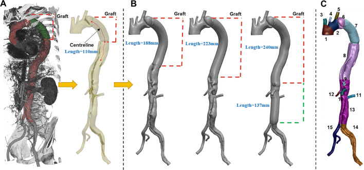
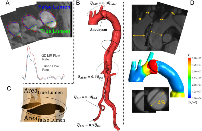
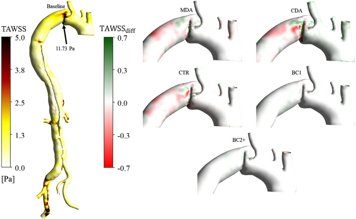
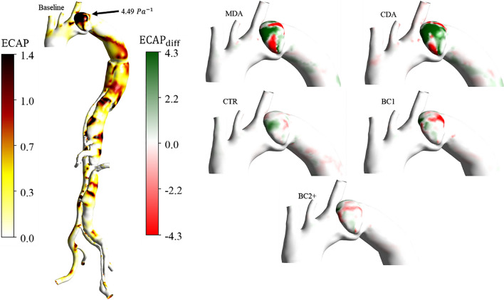
Materials and methods: A post-operative case of TBAD was segmented from computed tomography angiography data. Three virtual surgeries were generated using different grafts; two additional cases with compliant grafts were assessed. Compliant CFD simulations were performed using a patient-specific inlet flow rate and three-element Windkessel outlet boundary conditions informed by 2D-Flow MRI data. The wall compliance was calibrated using Cine-MRI images. Pressure, wall shear stress (WSS) indices and energy loss (EL) were computed.

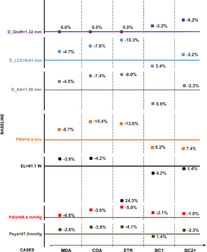
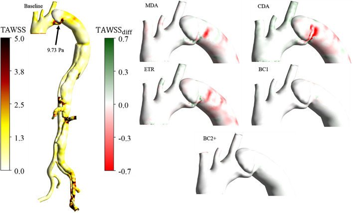
Results: Increased aortic stiffness and longer grafts increased aortic pressure and EL. Implementing a compliant graft matching the aortic compliance of the patient reduced the pulse pressure by 11% and EL by 4%. The endothelial cell activation potential (ECAP) differed the most within the aneurysm, where the maximum percentage difference between the reference case and the mid (MDA) and complete (CDA) descending aorta replacements increased by 16% and 20%, respectively.

Conclusion: This study suggests that by minimising graft length and matching its compliance to the native aorta whilst aligning with surgical requirements, the risk of LV hypertrophy may be reduced. This provides evidence that compliance-matching grafts may enhance patient outcomes.

Keywords: CFD simulation; Compliance mismatch; Dacron graft; Type-B aortic dissection; Virtual interventions.


Figures

Fig. 1
Fig. 1
A Automatic 3D rendering of the CT angiography, showing, in red, the aortic vessel and, in green, the graft; the segmented post-operative geometry resulting from the CT angiography is shown next to it-namely the baseline case. B The three virtual surgical scenarios created from the baseline case by varying the length of graft. The red centreline from which the grafts have been swept is shown on the post-operative geometry. Red and green dashed lines indicate the extent of the 32 mm and 28 mm diameter thoracoabdominal graft of ETR, respectively. The length of each graft is indicated in blue. C aortic regions defined along the centreline using anatomical landmarks to account for proximal variations of stiffness. The numbers indicate: 1 ascending aorta, 2 arch, 3 brachiocephalic trunk, 4 left common carotid, 5 left subclavian, 6 isthmus, 7 graft, 8 descending aorta, 9 coeliac trunk, 10 superior mesenteric artery, 11 left renal, 12 right renal, 13 abdominal aorta, 14 left iliac, 15 right iliac
Fig. 2
Fig. 2
A 2D-Flow MRI plane showing in blue and green the false and true lumen respectively; below are the extracted raw and rescaled by 30% flow rates, B Flow split at the outlets: 30% of the flow leaves through the supra-aortic branches, and 40% of the remaining flow leaves through the abdominal arteries following the true and false lumen shown in C. The remaining abdominal false and true lumen flows are split as 70/30% between the exterior and interior iliac arteries; the right exterior (REI) and interior (RII) iliac arteries are shown as an example. D Sample cine-MRI planes used to measure the stiffness of the aorta. The aortic arch of BC1 is zoomed in to show the distribution of local specific stiffness values K obtained for the case of a compliant graft
Fig 3
Fig 3
Y-axis represents systolic and diastolic pressures (Psys, Pdia), EL, PWV, and maximum diameter variation at the ascending aorta (D_AA), left common carotid (D_LCC), and graft (D_Graft) for the baseline case. The X-axis shows values of the metrics of interest for five additional cases, each labelled with their respective relative signed errors. Bold lines denote the metric values for the baseline case and those corresponding to 0% relative signed errors
Fig 4
Fig 4
Front view of the TAWSS. On the left, values over 5 Pa are found at the PET, the abdominal arteries, and the left iliac of the baseline case. The black arrow indicates the maximum TAWSS at the PET. On the right, the TAWSS differences between the baseline and the five cases are shown. A zoom is made on the AA and aortic arch as regions of interest where the TAWSS is high
Fig 5
Fig 5
Front view of the TAWSS. On the left, values over 5 Pa are found at the PET and sutures with the graft of the baseline case. The black arrow indicates the maximum TAWSS at the PET. On the right, the TAWSS differences between the baseline and the five other cases are shown. A zoom is made on the AA and aortic arch as regions of interest where the TAWSS is high
Fig 6
Fig 6
ECAP distributions, front view. On the left, ECAP absolute values for baseline case; values over 1.4 Pa-1 are noted in the aneurysm. The black arrow indicates the maximum ECAP value in the aneurysm. On the right, ECAP differences between the baseline and the five virtual cases

References

    1. Trahanas JM, Jarral OA, Long C, Hughes GC. Management of chronic type B aortic dissection. Vessel Plus. 2022 doi: 10.20517/2574-1209.2021.125. - DOI
    1. Williams ML, et al. Thoracic endovascular repair of chronic type B aortic dissection: a systematic review. Ann. Cardiothorac. Surg. 2022;11(1):1–15. doi: 10.21037/ACS-2021-TAES-25. - DOI - PMC - PubMed
    1. Hsieh RW, et al. Comparison of type B dissection by open, endovascular, and medical treatments. J. Vasc. Surg. 2019;70(6):1792–1800.e3. doi: 10.1016/j.jvs.2019.02.062. - DOI - PubMed
    1. Goodney PP, et al. Survival after open versus endovascular thoracic aortic aneurysm repair in an observational study of the medicare population. Cardiovasc. Surg. 2011 doi: 10.1161/CIRCULATIONAHA.111.033944/-/DC1. - DOI - PMC - PubMed
    1. Tian DH, De Silva RP, Wang T, Yan TD. Open surgical repair for chronic type B aortic dissection: a systematic review systematic review background. Ann. Cardiothorac. Surg. 2014;3(4):340–350. doi: 10.3978/j.issn.2225-319X.2014.07.10. - DOI - PMC - PubMed

MeSH terms

Substances

LinkOut - more resources