

From CNN to Transformer: A Review of Medical Image Segmentation Models
- PMID: 38438696
- PMCID: PMC11300773
- DOI: 10.1007/s10278-024-00981-7
From CNN to Transformer: A Review of Medical Image Segmentation Models
Abstract
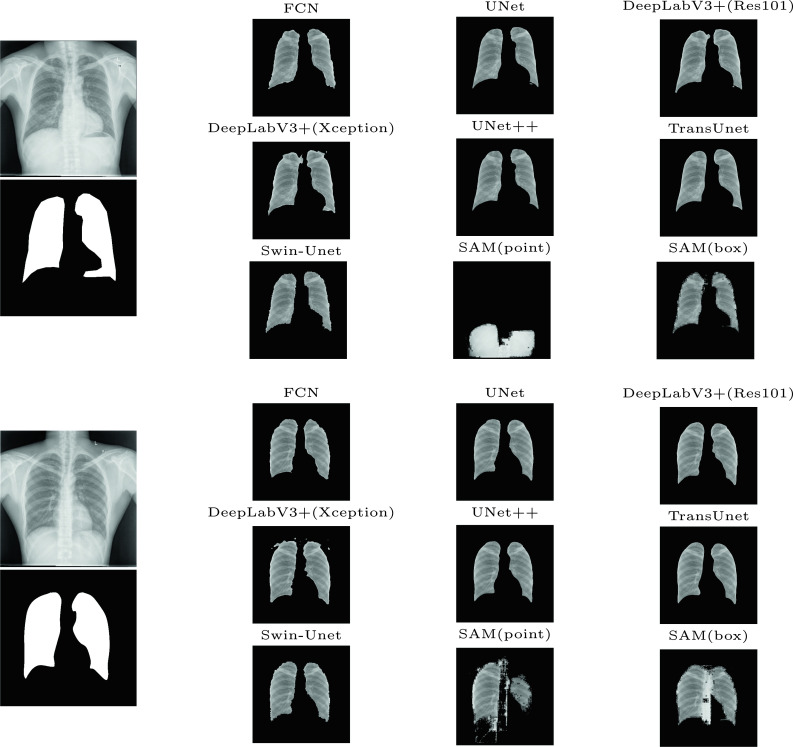
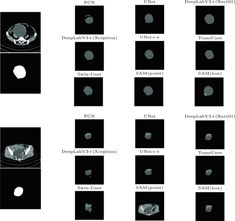
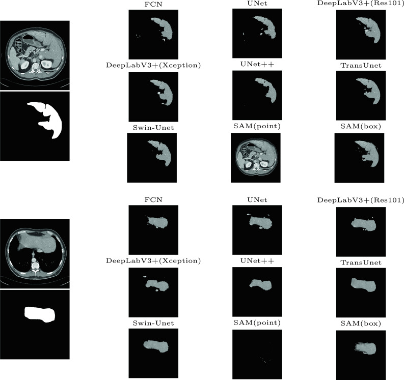
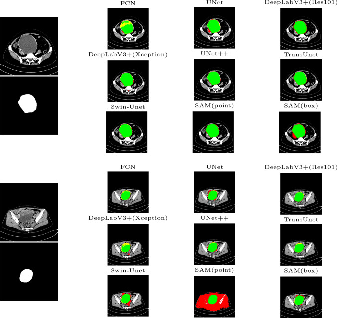
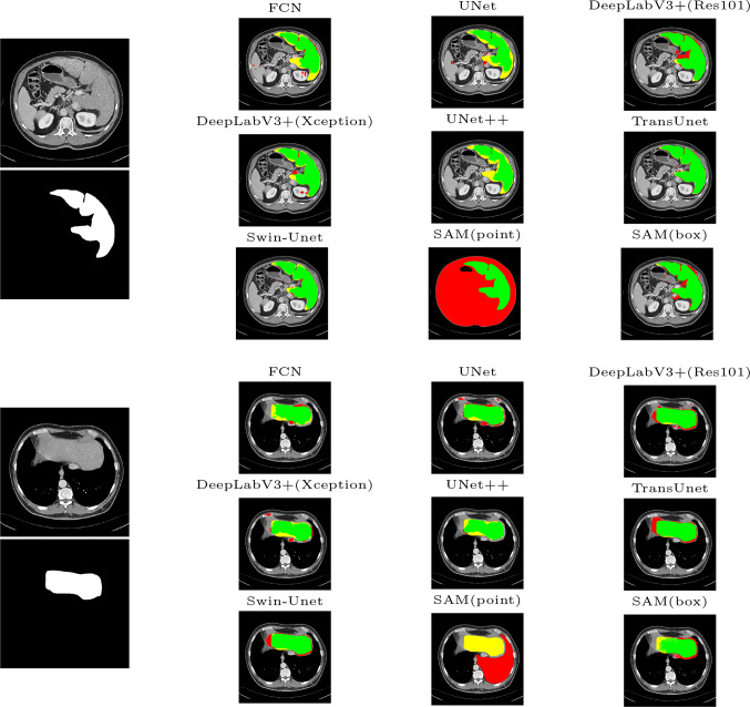
Medical image segmentation is an important step in medical image analysis, especially as a crucial prerequisite for efficient disease diagnosis and treatment. The use of deep learning for image segmentation has become a prevalent trend. The widely adopted approach currently is U-Net and its variants. Moreover, with the remarkable success of pre-trained models in natural language processing tasks, transformer-based models like TransUNet have achieved desirable performance on multiple medical image segmentation datasets. Recently, the Segment Anything Model (SAM) and its variants have also been attempted for medical image segmentation. In this paper, we conduct a survey of the most representative seven medical image segmentation models in recent years. We theoretically analyze the characteristics of these models and quantitatively evaluate their performance on Tuberculosis Chest X-rays, Ovarian Tumors, and Liver Segmentation datasets. Finally, we discuss the main challenges and future trends in medical image segmentation. Our work can assist researchers in the related field to quickly establish medical segmentation models tailored to specific regions.
Keywords: CNN; Deep learning; Medical image segmentation; Transformer; U-Net.
© 2024. The Author(s) under exclusive licence to Society for Imaging Informatics in Medicine.
Conflict of interest statement
The authors declare no competing interests.
Figures


References
-
- Golan, R., Jacob, C., Denzinger, J.: Lung nodule detection in ct images using deep convolutional neural networks. In: International Joint Conference on Neural Networks (2016)
-
- Christ, P.F., Ettlinger, F., Grün, F., Elshaera, M.E.A., Lipkova, J., Schlecht, S., Ahmaddy, F., Tatavarty, S., Bickel, M., Bilic, P.: Automatic liver and tumor segmentation of CT and MRI volumes using cascaded fully convolutional neural networks (2017)
-
- Otsu, N.: A threshold selection method from gray-level histograms. IEEE Transactions on Systems, Man, and Cybernetics 9(1), 62–66 (1979)
-
- Magnier, Baptiste: Edge detection: a review of dissimilarity evaluations and a proposed normalized measure. Multimedia Tools & Applications (2017)
Publication types
MeSH terms
Grants and funding
LinkOut - more resources
Full Text Sources