. 2024 Apr;25(4):1936-1961.
doi: 10.1038/s44319-024-00107-8. Epub 2024 Mar 4.

Identification of plants' functional counterpart of the metazoan mediator of DNA Damage checkpoint 1

Affiliations

Identification of plants' functional counterpart of the metazoan mediator of DNA Damage checkpoint 1

Zdravko J Lorković et al. EMBO Rep. 2024 Apr.

Abstract

Induction of DNA damage triggers rapid phosphorylation of the histone H2A.X (γH2A.X). In animals, mediator of DNA damage checkpoint 1 (MDC1) binds γH2A.X through a tandem BRCA1 carboxyl-terminal (tBRCT) domain and mediates recruitment of downstream effectors of DNA damage response (DDR). However, readers of this modification in plants have remained elusive. We show that from the Arabidopsis BRCT domain proteome, BCP1-4 proteins with tBRCT domains are involved in DDR. Through its tBRCT domain BCP4 binds γH2A.X in vitro and localizes to DNA damage-induced foci in an H2A.X-dependent manner. BCP4 also contains a domain that interacts directly with NBS1 and thus acts as a functional counterpart of MDC1. We also show that BCP1, that contains two tBRCT domains, co-localizes with γH2A.X but it does not bind γH2A.X suggesting functional similarity with human PAXIP1. A phylogenetic analysis supports that PAXIP1 and MDC1 in metazoa and their plant counterparts evolved independently from common ancestors with tBRCT domains. Collectively, our study reveals missing components and provides mechanistic and evolutionary insights into plant DDR.

Keywords: BCP4; BRCT Domain; DNA Damage Response; Histone H2A.X; MDC1.


Conflict of interest statement

The authors declare no competing interests.

Figures
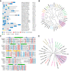
Figure 1
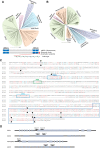
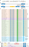
Figure 1. Identification of Arabidopsis proteins with BRCT domains.
(A) Schematic representation of 21 Arabidopsis proteins containing BRCT domains. The gene codes, protein names, and length in amino acids (aa) are indicated. (B) Human and Arabidopsis BRCT domain proteins were aligned with MUSCLE and a maximum likelihood circular cladogram was generated with CLC Genomics Workbench ver. 11.0. (C) Sequence alignment of Arabidopsis BCP3 and BCP4 and human MDC1 and PAXIP1 tBRCT domains. For alignment, tBRCT3 of PAXIP1 was used. Residues conserved in at least four proteins are shaded light blue and those similar in at least four proteins are shaded light red. Structural elements of domains are indicated on the top of the alignment according to Stucki et al (2005). Amino acids involved in MDC1 interaction with γH2A.X are indicated with red dots. (D) All human and Arabidopsis tBRCT domains were aligned as in (B) and a maximum likelihood tree illustrating amino acid sequence conservation between tBRCT domains was generated with CLC Genomics Workbench ver. 11.0. (B, D) Human and Arabidopsis proteins with high sequence homology share the same color code. Arabidopsis proteins and tBRCT domains clustering with human MDC1 and PAXIP1 are indicated in red and shaded in purple.
Figure 2
Figure 2. Analysis of bcp mutants.
(A) DNA damage sensitivity of BCP mutant lines assessed by true leaf development assay (n = 13 biological replicates, each with 64 seeds). DNA damage-sensitive mutants of H2A.X (hta3 hta5) and H2A.W.7 (hta7) histone variants were used as controls. Data information: Data are represented as a box plot with median and interquartile range (box) and minimal and maximal values (whiskers). ***P  ≤  0.0001 (two-tailed paired Student’s t test). (B) Analysis of γH2A.X levels in bcp mutant seedlings after induction of DNA damage for 2 h. Representative western blots for γH2A.X and H2A.X in BCP mutants. Quantified γH2A.X levels normalized to the total H2A.X (n = 6, three biological replicates with two technical repeats). (C) Analysis of γH2A.X foci in nuclei of WT and bcp mutants. Data information: Maximum intensity projection images from Z-stacks of representative nuclei are shown. Scale bars represent 5 µm. (D) Quantification of γH2A.X foci number in bcp mutants. For each genetic background n = 8 nuclei were used. Data are represented as a box plot with median and interquartile range (box) and minimal and maximal values (whiskers). Data information: P  >  0.05, ns (nonsignificant). (two-tailed paired Student’s t test). (E) DNA damage sensitivity of BCP-mClover3 complementing lines (n = 6 biological replicates, each with 64 seeds). Data information: In (B, E), data are presented as mean ± SD. ***P ≤ 0.0001; **P ≤ 0.001; *P ≤ 0.01; P > 0.01, ns (nonsignificant). (two-tailed paired Student’s t test). In (A, E) Seeds were germinated on medium containing 20 μg/ml of zeocin and true leaf development was scored 12 days after germination. Source data are available online for this figure.
Figure 3
Figure 3. BCP4 and MDC1 share similar properties.
(A) BCP1 and BCP4 co-localize with γH2A.X foci. Immunostaining of nuclei from bleomycin-treated seedlings expressing BCP1-mClover3 and BCP4-mClover3 in bcp1 and bcp4 mutants, respectively. (B, C) BCP1 and BCP4 foci formation is dependent on H2A.X Immunostaining of nuclei from bleomycin-treated seedlings expressing BCP1-mClover (B) and BCP4-mClover (C) in bcp1 hta3 hta5 and bcp4 hta3 hta5 genetic backgrounds, respectively. (D) BCP4 binds phosphorylated H2A.X C-terminal peptide. Affinity pull-down with N- and C-terminally His-tagged BCP4 and biotinylated H2A.X peptides. (E) Affinity pull-down with GST-tagged tBRCT domain of BCP4. (F) The tBRCT domains of BCP1 do not bind phosphorylated H2A.X C-terminal peptide. Affinity pull-down with GST-tagged tBRCT domains and biotinylated H2A.X peptides. Data information: In (AC), maximum intensity projection images from Z-stacks are shown and scale bars represent 5 µm. Proteins were analyzed on 10% (D) or 12% (E, F) SDS-PAGE and gels were stained with Coomassie blue. Source data are available online for this figure.
Figure 4
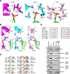
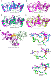
Figure 4. BCP4 binds γH2A.X through a structurally conserved binding pocket.
(A) Comparison of interactions of tBRCT domains of MDC1, BCP4, and BCP3 with phosphorylated C-terminal peptide of H2A.X. Published (MDC1; Stucki et al, 2005) and AlphaFold2 predicted (BCP3 and BCP4) contacts of indicated amino acids with pSer of H2A.X are indicated with dotted lines. (B) Contacts of the human Arg1933 with the C-terminal carboxyl group of H2A.X and Glu2063 are conserved in Arabidopsis BCP3 and BCP4. (C) Phosphoserine-binding consensus sequences of BCP3, BCP4, and MDC1. The coloring of amino acids is according to their polarity. (D) Sequence alignment of MDC1 and BCP4 point mutants comprising γH2A.X-binding site. MDC1 and BCP4 amino acids involved and predicted in binding of γH2A.X are indicated in red (top panel). Phosphopeptide pull-down analysis of wild-type and mutant GST-tBRCT domain of BCP4 (bottom panels). GST-tBRCT concentrations used with phosphopeptide were 2.5, and 20 mg/ml, respectively. Input lanes were loaded with 1.5 µg. Proteins were analyzed on 12% SDS-PAGE and gels were stained with Coomassie blue. Source data are available online for this figure.
Figure 5
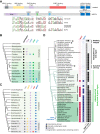
Figure 5. Comparison of BCP4 and MDC1 sequence motifs.
(A) Schematic representation of human MDC1 and Arabidopsis BCP4 proteins. Conserved sequence motifs are indicated, and consensus sequences of plant motifs are depicted at the bottom. (B) Summary of the presence/absence of BCP3/BCP4 sequence motifs across Viridiplantae. Open circles denote phylogenetic groups where motif is present in only a subset of analyzed species. (C) In metazoa, functional motifs identified in human MDC1 are unique to mammals. The numbers of MDC1 proteins from each phylogenetic group used for the creation of cladogram are indicated in parentheses. (D) Evolutionary trajectories of BCP1, BCP4, and H2A.X in Archaeplastida. Protein presence is displayed at the tip of each branch, and major groups are denoted next to phylogeny. For H2A.X, (i) the presence of H2A, (ii) SQEF or SQEF-like motifs, and (iii) the presence of a monophyletic clade containing Arabidopsis H2A.X (Fig. EV4) were displayed separately. Data information: Plant and metazoan protein sequences used for the analysis shown in A–C are available in Source data. Source data are available online for this figure.
Figure 6
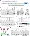
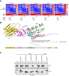
Figure 6. BCP4 interacts with NBS1 of the MRN complex.
(A) Schematic presentation of BCP4 with sequences of TQxϕ and SQ/DWD sequences used for protein expression. (B) Co-purification of full-length NBS1 and the FHA+tBRCT domains of NBS1 with GST and GST-tagged TQxϕ or SQ/DWD motifs of BCP4. (C) GST pull-down with independently purified recombinant proteins. Coomassie-stained gels of purified proteins are on two left panels. (D) Co-purification of His6- FHA+tBRCT of NBS1 with the SQ/DWD deletion mutant forms of BCP4. (E) Schematic presentation of BCP4 SQ/DWD deletion mutant forms and summary of interactions with NBS1. (F) Sequence alignment of the SQ/DWD motifs of BCP3 and BCP4. Identical and similar amino acids are highlighted on black and gray background, respectively. Blue background indicates amino acids predicted to interact with NBS1. (G) AlphaFold2 predicted interaction of NBS1 and the SQ/DWD motif of BCP4. Amino acids predicted to interact are indicated with numbers corresponding to full-length proteins. (H) Co-purification of full-length NBS1 or the FHA+tBRCT domains of NBS1 with GST-tagged deletion 4 of BCP4 SQ/DWD motif and its point mutants as indicated. Source data are available online for this figure.
Figure EV1
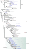
Figure EV1. Phylogeny of BCP1, BCP3, and BCP4.
(A) Maximum likelihood tree of BCP1 across Viridiplantae. The schematic presentation of BCP1 is shown at the bottom and a PHD finger present in all BCP sequences except in Brassicaceae is indicated. (B) Maximum likelihood tree of BCP3 and BCP4 across Viridiplantae. (A, B) Major clades are indicated by differently colored shading. The non-flowering land plant clade includes sequences from hornworts, mosses, liverworts, lycophytes, and monilophytes. Thuja plicata was the only gymnosperm used in the analysis. (C) Alignment of BCP3 and BCP4 proteins. Identical and conserved amino acids are indicated in red and blue letters, respectively. The positions of introns are indicated by black arrows. The tBRCT domain and three other conserved regions are shown in colored boxes. (D) Schematic representation of Arabidopsis BCP genes with exons indicated by gray boxes and introns by black lines. Exons and introns are drawn to scale according to the lengths of DNA sequences. Positions of T-DNA insertions in bcp mutant lines used for DNA damage sensitivity assays are indicated above each gene. Data information: A list of protein sequences used for the analysis is available from Source data for Fig. 5A,B.
Figure EV2
Figure EV2. Primary sequences analysis of BCP proteins.
(A) The consensus sequence of the PHD finger from BCP1 derived from the alignment of plant BCP1 proteins. Cysteine and histidine residues characteristic of PHD fingers are underlined. (B) Schematic representation of Arabidopsis BCP1 and human PAXIP1 proteins. Conserved domains and motifs of PAXIP1 and their assigned functions are indicated. A list of plant species and the corresponding protein sequences used for the analysis in (A, B) are available from Source data. (C) Alignment of SQSQ, DWD, and DTQ sequence motifs from BCP4. The signature motifs are shaded in blue and green. (D) Schematic representation of MDC1 proteins from invertebrate, vertebrate (except mammals), and mammalian species. Conserved domains and motifs and their assigned functions are indicated. Source data are available online for this figure.
Figure EV3
Figure EV3. Predicted structures of BCP proteins.
(AE) AlphaFold2 models of the tBRCT domains of BCP4 (A), BCP3 (B), BCP2 (C), and BCP1 (D, E) superimposed with a structure of human MDC1 in complex with the phosphorylated C-terminal peptide of H2A.X (Stucki et al, 2005). Note that tBRCT1 of BCP1 overlaps only with MDC1 N-terminal BRCT domain. This is presumably due to the absence of alpha helices (αL1 and αL2, in orange) in tBRCT1 of BCP1 connecting two BRCT domains as indicated in orange. In all panels, human MDC1 tBRCT is purple colored. (F) Comparison of interactions of the tBRCT domains of BCP1 and BCP2 with phosphorylated C-terminal peptide of H2A.X. Published (MDC1; Stucki et al, 2005) and AlphaFold2 predicted (BCP1 and BCP2) contacts of amino acids with pSer of H2A.X are indicated respectively with green and orange dotted lines. For a comparison, BCP4 is displayed to indicate reduced abilities of the tBRCT domains of BCP1 and BCP2 to contact pSer of H2A.X. Human MDC1 tBRCT is purple colored.
Figure EV4
Figure EV4. Phylogeny of H2A variants from Archaeplastida H2A orthogroup.
The phylogenetic positions of Arabidopsis H2A variants are marked in red. H2A.X sequences with a SQEF/Y motif or SQEF-like (SQ + E/D + F/I/L/V/Y) motif at the C-terminus are highlighted in blue. Note that in green algae, red algae, and glaucophytes H2A and SQEF/Y motif-containing H2As do not form separate clades.
Figure EV5
Figure EV5. Predicted structure of NBS1 and its interaction with SQ/DWD region of BCP4.
(A) PAE plots of NBS1 (1–324) and BCP4 SQ/DWD (276–325), with calculated predicted local distance difference test (pLDDT), predicted template modeling (PTM), and interface-predicted template modeling (iPTM) scores. (B) AlphaFold2 model of Arabidopsis NBS1 in complex with SQ/DWD region of BCP4 (top panel). A sequence of BCP4 minimal peptide interacting with NBS1 is indicated with Leu and Cys residues involved in interaction with NBS1 highlighted in red. Schematic presentation of NBS1 with indicated conserved domains (bottom panel). (C) Co-purification of NBS1 with GST-tagged deletion 4 of BCP4 SQ/DWD motif and its point mutants. Source data are available online for this figure.

Similar articles

Cited by

References

    1. Akutsu N, Iijima K, Hinata T, Tauchi H. Characterization of the plant homolog of Nijmegen breakage syndrome 1: Involvement in DNA repair and recombination. Biochem Biophys Res Commun. 2007;353:394–408. doi: 10.1016/j.bbrc.2006.12.030. - DOI - PubMed
    1. Almawi AW, Matthews LA, Guarne A. FHA domains: phosphopeptide binding and beyond. Prog Biophys Mol Biol. 2017;127:105–110. doi: 10.1016/j.pbiomolbio.2016.12.003. - DOI - PubMed
    1. Buchfink B, Xie C, Huson DH. Fast and sensitive protein alignment using DIAMOND. Nat Methods. 2015;12:59–60. doi: 10.1038/nmeth.3176. - DOI - PubMed
    1. Callebaut I, Mornon PJ. From BRCA1 to RAP1: a widespread BRCT module closely associated with DNA repair. FEBS Lett. 1997;400:25–30. doi: 10.1016/S0014-5793(96)01312-9. - DOI - PubMed
    1. Chapman JR, Jackson PS. Phospho-dependent interactions between NBS1 and MDC1 mediate chromatin retention of the MRN complex at sites of DNA damage. EMBO Rep. 2008;9:795–801. doi: 10.1038/embor.2008.103. - DOI - PMC - PubMed