

Shallow and deep learning classifiers in medical image analysis
- PMID: 38438821
- PMCID: PMC10912073
- DOI: 10.1186/s41747-024-00428-2
Shallow and deep learning classifiers in medical image analysis
Abstract
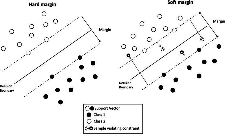
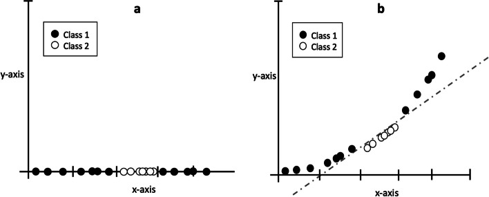
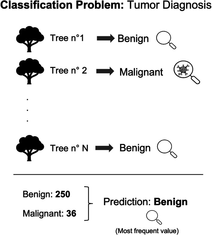
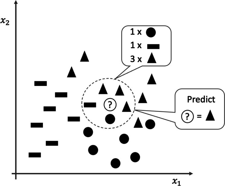
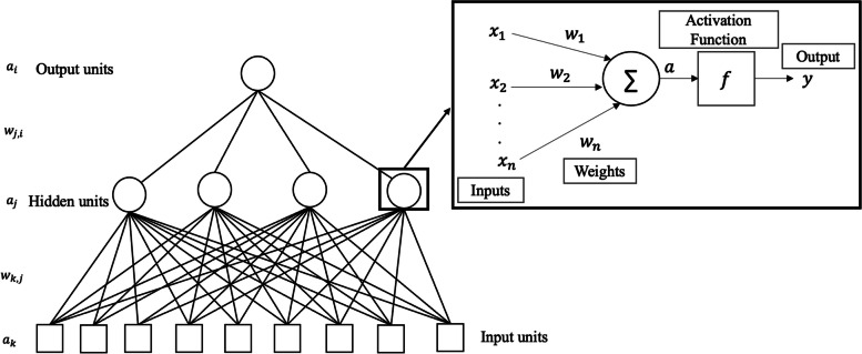
An increasingly strong connection between artificial intelligence and medicine has enabled the development of predictive models capable of supporting physicians' decision-making. Artificial intelligence encompasses much more than machine learning, which nevertheless is its most cited and used sub-branch in the last decade. Since most clinical problems can be modeled through machine learning classifiers, it is essential to discuss their main elements. This review aims to give primary educational insights on the most accessible and widely employed classifiers in radiology field, distinguishing between "shallow" learning (i.e., traditional machine learning) algorithms, including support vector machines, random forest and XGBoost, and "deep" learning architectures including convolutional neural networks and vision transformers. In addition, the paper outlines the key steps for classifiers training and highlights the differences between the most common algorithms and architectures. Although the choice of an algorithm depends on the task and dataset dealing with, general guidelines for classifier selection are proposed in relation to task analysis, dataset size, explainability requirements, and available computing resources. Considering the enormous interest in these innovative models and architectures, the problem of machine learning algorithms interpretability is finally discussed, providing a future perspective on trustworthy artificial intelligence.Relevance statement The growing synergy between artificial intelligence and medicine fosters predictive models aiding physicians. Machine learning classifiers, from shallow learning to deep learning, are offering crucial insights for the development of clinical decision support systems in healthcare. Explainability is a key feature of models that leads systems toward integration into clinical practice. Key points • Training a shallow classifier requires extracting disease-related features from region of interests (e.g., radiomics).• Deep classifiers implement automatic feature extraction and classification.• The classifier selection is based on data and computational resources availability, task, and explanation needs.
Keywords: Artificial intelligence; Deep learning; Explainable AI; Machine learning classifiers; Shallow learning.
© 2024. The Author(s).
Conflict of interest statement
The authors declare that they have no competing interests.
Figures
Similar articles
-
Artificial intelligence: Deep learning in oncological radiomics and challenges of interpretability and data harmonization.Phys Med. 2021 Mar;83:108-121. doi: 10.1016/j.ejmp.2021.03.009. Epub 2021 Mar 22. Phys Med. 2021. PMID: 33765601 Review.
-
Image biomarkers and explainable AI: handcrafted features versus deep learned features.Eur Radiol Exp. 2024 Nov 19;8(1):130. doi: 10.1186/s41747-024-00529-y. Eur Radiol Exp. 2024. PMID: 39560820 Free PMC article. Review.
-
Applications of and issues with machine learning in medicine: Bridging the gap with explainable AI.Biosci Trends. 2025 Jan 14;18(6):497-504. doi: 10.5582/bst.2024.01342. Epub 2024 Dec 8. Biosci Trends. 2025. PMID: 39647859 Review.
-
Data Integration Using Advances in Machine Learning in Drug Discovery and Molecular Biology.Methods Mol Biol. 2021;2190:167-184. doi: 10.1007/978-1-0716-0826-5_7. Methods Mol Biol. 2021. PMID: 32804365 Review.
-
Explainable AI for Intraoperative Motor-Evoked Potential Muscle Classification in Neurosurgery: Bicentric Retrospective Study.J Med Internet Res. 2025 Mar 24;27:e63937. doi: 10.2196/63937. J Med Internet Res. 2025. PMID: 40127441 Free PMC article.
Cited by
-
Machine learning in shoulder arthroplasty : a systematic review of predictive analytics applications.Bone Jt Open. 2025 Feb 4;6(2):126-134. doi: 10.1302/2633-1462.62.BJO-2024-0234.R1. Bone Jt Open. 2025. PMID: 39900101 Free PMC article.
References
Publication types
MeSH terms
Grants and funding
LinkOut - more resources
Full Text Sources