. 2024 Mar 4;25(1):6.
doi: 10.1186/s12860-024-00502-y.

TonEBP/NFAT5 expression is associated with cisplatin resistance and migration in macrophage-induced A549 cells

Affiliations

TonEBP/NFAT5 expression is associated with cisplatin resistance and migration in macrophage-induced A549 cells

Hee Ju Song et al. BMC Mol Cell Biol. .

Abstract

Background: Macrophages promote angiogenesis, metastasis, and drug resistance in several cancers. Similarly, TonEBP/NFAT5 induces metastasis in renal carcinoma and colon cancer cells. However, the role of this transcription factor and that of macrophages in lung cancer cells remains unclear. Therefore, this study investigated the effects of macrophages and TonEBP/NFAT5 expression on cisplatin resistance and migration in A549 lung adenocarcinoma cells.

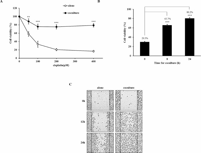
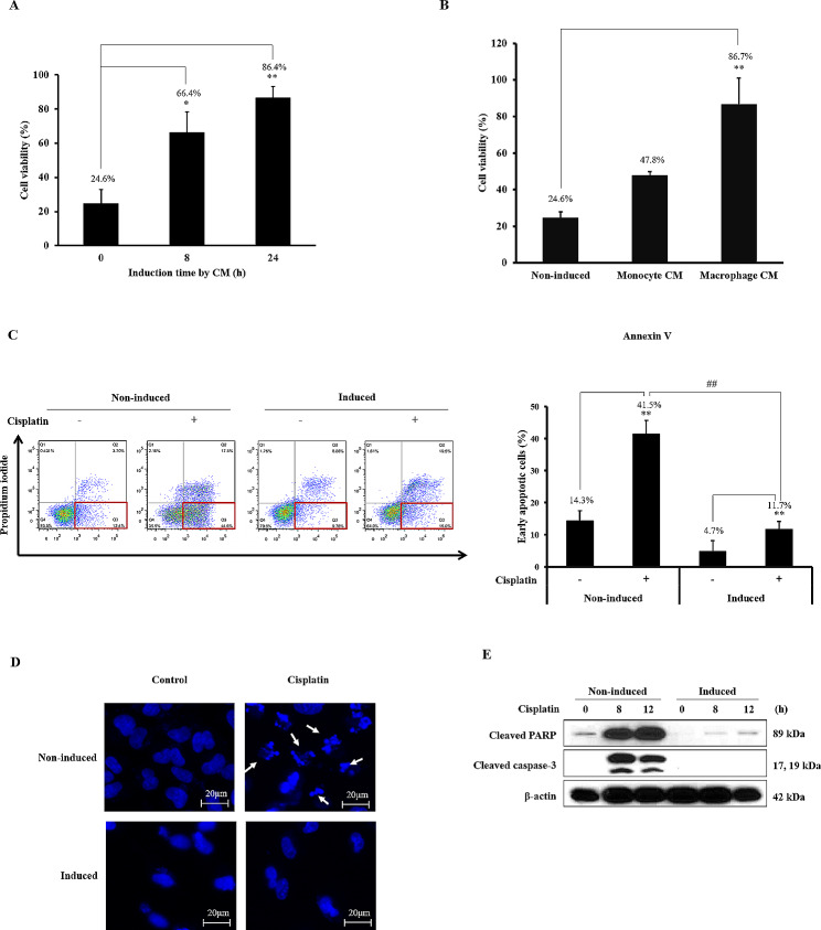
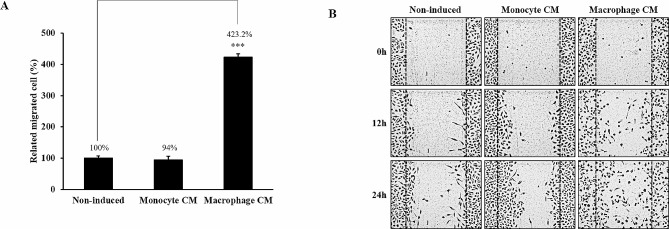
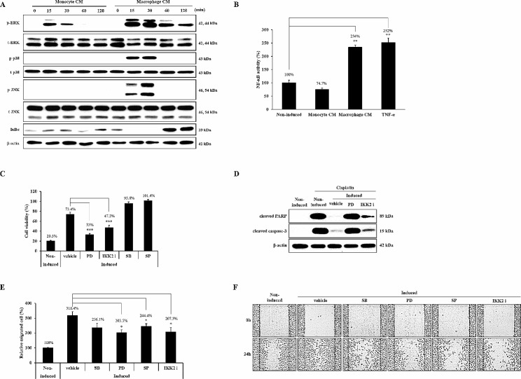
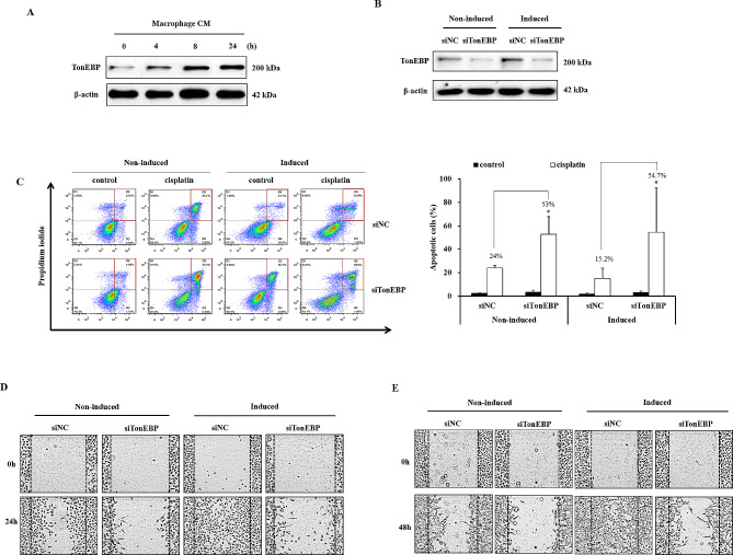
Results: A549 cells were cultured alone or indirectly co-cultured with THP-1-derived macrophages using a transwell culture chamber. Cisplatin-induced cell death was markedly decreased and migration increased in co-cultured A549 cells. Macrophage-conditioned media (CM) showed a similar effect on drug resistance and migration. Cisplatin-induced apoptosis, DNA fragmentation, and cleaved apoptotic proteins PARP and caspase-3 were markedly reduced in macrophage CM-induced A549 cells. Here, ERK, p38, JNK, and NF-κB activities were increased by macrophage CM. Furthermore, the proteins involved in cisplatin resistance and cancer cell migration were identified using specific inhibitors of each protein. ERK and NF-κB inhibition considerably reduced cisplatin resistance. The increase in macrophage CM-induced migration was partially reduced by treatment with ERK, JNK, and NF-κB inhibitors. TonEBP/NFAT5 expression was increased by macrophages, resulting in increased cisplatin resistance, cell migration, and invasion. Moreover, RNAi-mediated knockdown of TonEBP/NFAT5 reduced cisplatin resistance, migration, and invasion in macrophage CM-induced A549 cells.

Conclusions: These findings demonstrate that paracrine factors secreted from macrophages can change A549 cells, resulting in the induction of drug resistance against cisplatin and migration. In addition, the TonEBP/NFAT5 ratio, increased by macrophages, is an important regulator of the malignant transformation of cells.

Keywords: A549; Cisplatin resistance; Migration; TonEBP/NFAT5.


Conflict of interest statement

The authors declare no competing interests.

Figures

Fig. 1
Fig. 1
Effect of macrophage co-culture on cisplatin resistance and migration. (A) A549 cells were cultured alone or co-culutred with THP-1 derived macrophages for 24 h and then treated with various concentrations (0 ~ 400 µM) of cisplatin for 24 h. (B) A549 cells were cultured alone or co-culutred with THP-1 derived macrophages for 8 or 24 h and then treated with 200 µM cisplatin for 24 h. The cell viability was determined using MTT assay. (C) Migration ability was determined using a wound healing assay in A549 cells co-cultured with macrophages for 24 h. Data are average values ± SD of at least six independent experiments. ** p < 0.01, *** p < 0.001 (compared with alone and 0 h)
Fig. 2
Fig. 2
Induction of cisplatin resistance by macrophage CM. (A) A549 cells were incubated with macrophage conditioned media (CM) for 8 or 24 h and then treated with 200 µM cisplatin for 24 h. (B) A549 cells were incubated with monocyte or macrophage conditioned media (CM) for 24 h and then treated with 200 µM cisplatin for 24 h. Cell viability was evaluated using a MTT assay. (C-E) A549 cells were induced with macrophage CM and treated with 200 µM cisplatin for 8 h. Cisplatin induced early apoptotic cells were identified using Annexin V and PI staining. Cisplatin induced DNA fragmentation was identified using DAPI staining. Cleavage of apoptotic proteins, PARP and caspase-3 were determined by immunoblotting. Full-length blots are presented in Supplementary Fig. [1]. Data are average values ± SD of at least three independent experiments. * p < 0.05, ** p < 0.01 (compared with 0 h and no cisplatin-treated group), ## p < 0.01 (compared with CM-induced cisplatin-treated group)
Fig. 3
Fig. 3
Induction of migration by macrophage CM. (A)A549 cells were incubated with monocyte or macrophage conditioned media (CM) for 24 h. Migration ability was determined using a wound healing assay. (B) Migrated cells were quantitated using a trans migration assay. Data are average values ± SD of at least five independent experiments. *** p < 0.001 (compared with Non-induced)
Fig. 4
Fig. 4
Macrophage CM induces cisplatin resistance and migration in NSCLC. (A) A549, Calu-3, H460, and H1299 cells were incubated with macrophage conditioned media (CM) for 24 h and then treated with 200 µM cisplatin for another 24 h. Cell viability was evaluated using a MTT assay. (B) Migrated cells were quantitated using a trans migration assay. Data are average values ± SD of at least three independent experiments. * p < 0.05, ** p < 0.01 (compared with Non-induced)
Fig. 5
Fig. 5
Signaling pathways are involved in macrophage-induced cisplatin resistance and migration. (A) A549 cells were incubated in monocyte or macrophage CM for the indicated times. Phosphorylation or the expression level of each protein was detected by western blotting using specific antibodies. The experiment was repeated with similar results. (B) A549 cells were transfected with an NF-κB-dependent luciferase expression plasmid and then incubated in CM or treated with 15 ng/ml TNF-α for 8 h. (C) A549 cells were induced with macrophage CM in the presence or absence of each inhibitor for 24 h, respectively. Viable cells in each condition were measured and compared with the corresponding control. (D) A549 cells were incubated with macrophage CM containing PD98059 or IKK2 inhibitor IV (IKK2 i). Cisplatin was added into each cell for 8 h. Whole cell lysates were subjected to western blotting. (E, F) A549 cells were treated with macrophage CM containing each inhibitor for 24 h. Migration ability was determined using a trans migration assay and a wound healing assay. The wound healing assay was repeated six times and representative images are shown. Full-length blots are presented in Supplementary Fig. (2). Data are average values ± SD of at least three independent experiments. * p < 0.05, ** p < 0.01, *** p < 0.001 (compared with Non-induced and vehicle)
Fig. 6
Fig. 6
Effect of TonEBP expression of cisplatin resistance, migration and invasion in macrophage-induced A549 cells. (A) A549 cells were incubated in macrophage CM for the indicated times. The expression of TonEBP was analyzed by immunoblotting. (B) A549 cells were transfected with negative control or TonEBP siRNA. TonEBP knocked down cells were incubated in macrophage CM for 24 h. (C) Apoptosis was measured by Annexin-V assay. TonEBP knocked down cells were treated with 200 µM cisplatin for 8 h. Annexin-V and PI-stained cells were analyzed using flow cytometry. (D-E) TonEBP knocked down cells were incubated in macrophage CM for 24 h. Migration ability was determined using wound healing assay. Invasion ability was measured by adding 200 µL Matrigel after wounding. Full-length blots are presented in Supplementary Fig. (3). Data are average values ± SD of at least three independent experiments. * p < 0.05, (compared with siNC)

Similar articles

Cited by

References

    1. Siegel R, Ma J, Zou Z, Jemal AJC. Cancer statistics, 2014. 2014;64(1):9–29. - PubMed
    1. Ceresoli GL, Cappuzzo F, Gregorc V, Bartolini S, Crino L, Villa EJAO. Gefitinib in patients with brain metastases from non-small-cell lung cancer: a prospective trial. 2004;15(7):1042–7. - PubMed
    1. Reck M, Popat S, Reinmuth N, De Ruysscher D, Kerr K, Peters SJA. Metastatic non-small-cell lung cancer (NSCLC): ESMO Clinical Practice guidelines for diagnosis, treatment and follow-up. 2014;25:iii27–iii39. - PubMed
    1. Wang G, Reed E, Li QQJO. Molecular basis of cellular response to cisplatin chemotherapy in non-small cell lung cancer. 2004;12(5):955–65. - PubMed
    1. Hinshaw DC, Shevde LAJC. The tumor microenvironment innately modulates cancer progression. 2019;79(18):4557–66. - PMC - PubMed