. 2024 Mar 4;13(1):13.
doi: 10.1186/s40035-024-00403-2.

LRRK2 kinase inhibition reverses G2019S mutation-dependent effects on tau pathology progression

Affiliations

LRRK2 kinase inhibition reverses G2019S mutation-dependent effects on tau pathology progression

Noah Lubben et al. Transl Neurodegener. .

Abstract

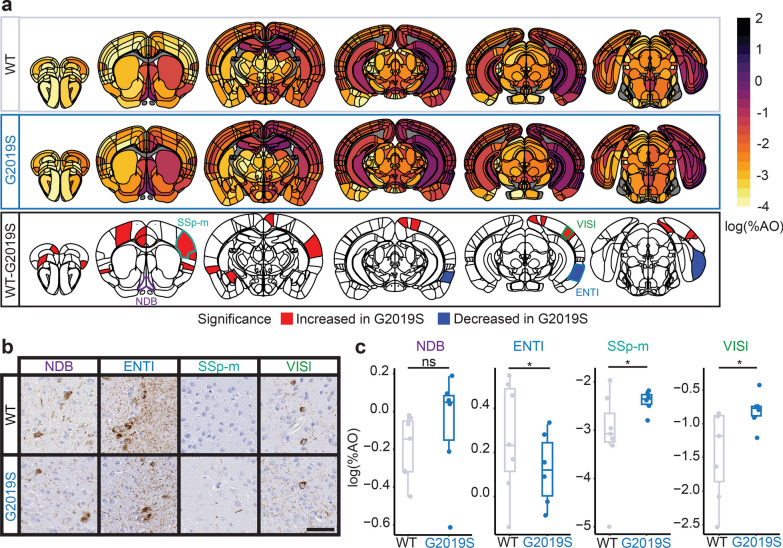
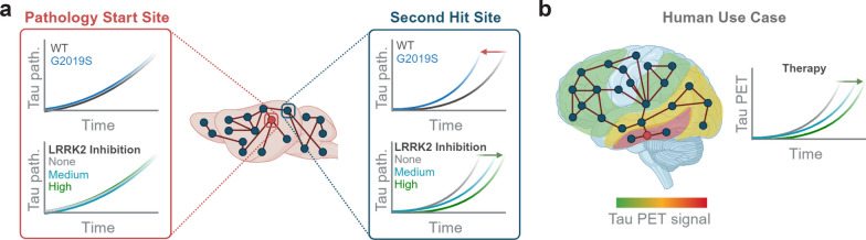
Background: Mutations in leucine-rich repeat kinase 2 (LRRK2) are the most common cause of familial Parkinson's disease (PD). These mutations elevate the LRRK2 kinase activity, making LRRK2 kinase inhibitors an attractive therapeutic. LRRK2 kinase activity has been consistently linked to specific cell signaling pathways, mostly related to organelle trafficking and homeostasis, but its relationship to PD pathogenesis has been more difficult to define. LRRK2-PD patients consistently present with loss of dopaminergic neurons in the substantia nigra but show variable development of Lewy body or tau tangle pathology. Animal models carrying LRRK2 mutations do not develop robust PD-related phenotypes spontaneously, hampering the assessment of the efficacy of LRRK2 inhibitors against disease processes. We hypothesized that mutations in LRRK2 may not be directly related to a single disease pathway, but instead may elevate the susceptibility to multiple disease processes, depending on the disease trigger. To test this hypothesis, we have previously evaluated progression of α-synuclein and tau pathologies following injection of proteopathic seeds. We demonstrated that transgenic mice overexpressing mutant LRRK2 show alterations in the brain-wide progression of pathology, especially at older ages.

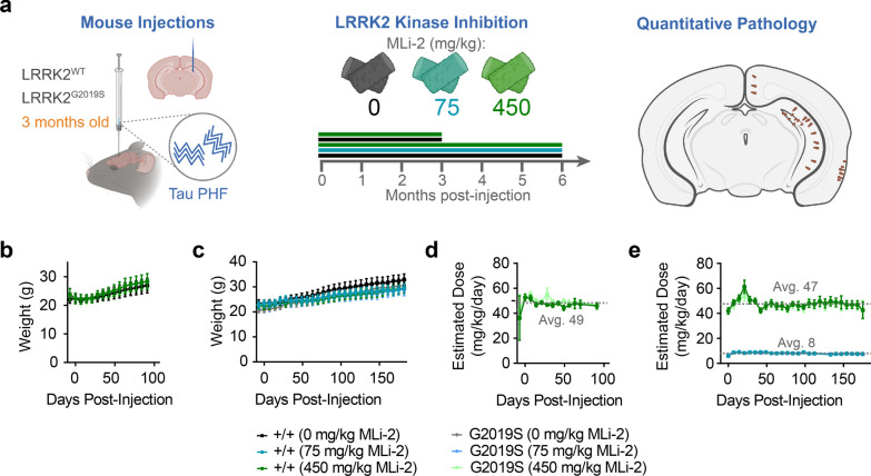
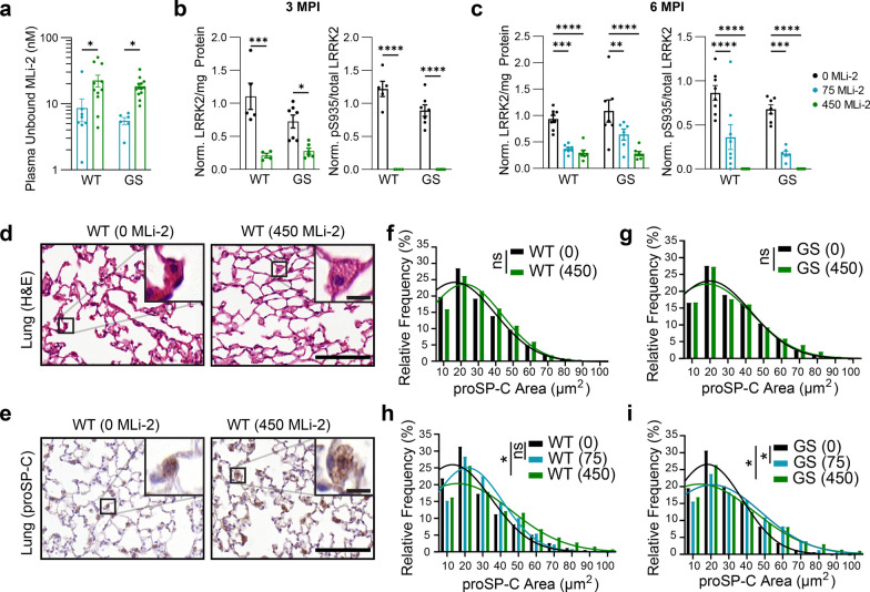
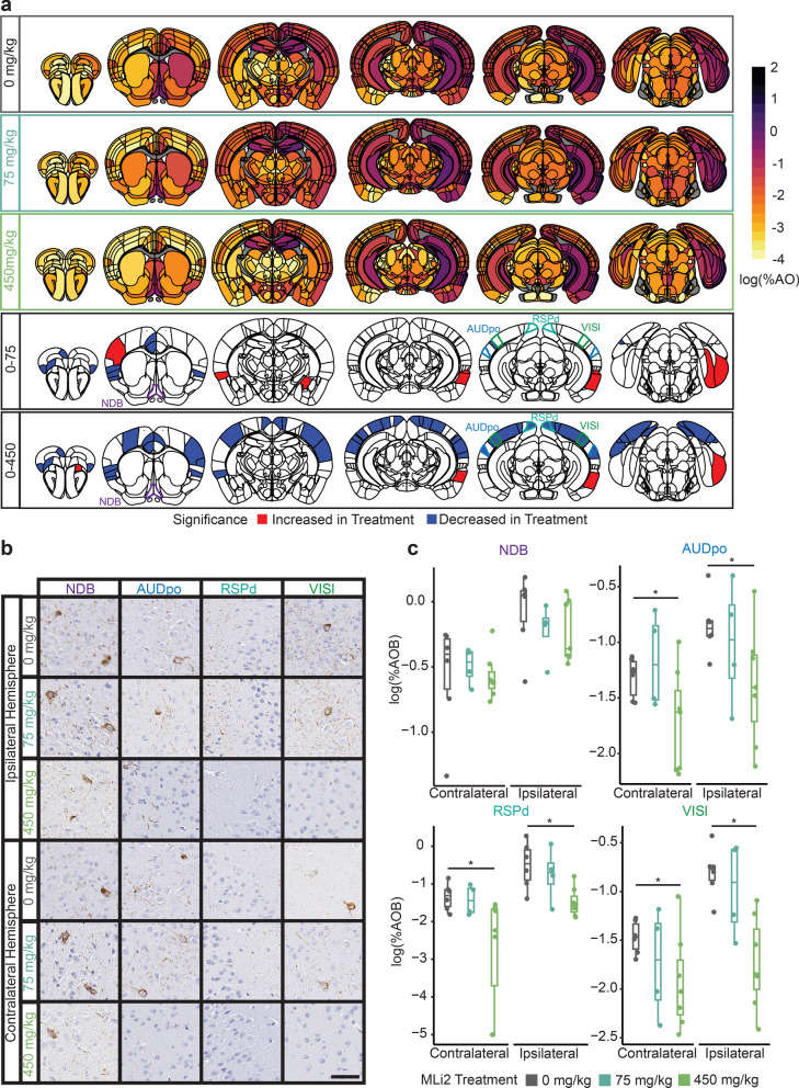
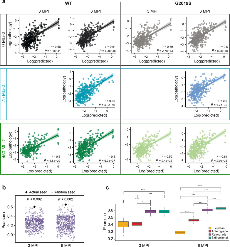
Methods: Here, we assess tau pathology progression in relation to long-term LRRK2 kinase inhibition. Wild-type or LRRK2G2019S knock-in mice were injected with tau fibrils and treated with control diet or diet containing LRRK2 kinase inhibitor MLi-2 targeting the IC50 or IC90 of LRRK2 for 3-6 months. Mice were evaluated for tau pathology by brain-wide quantitative pathology in 844 brain regions and subsequent linear diffusion modeling of progression.

Results: Consistent with our previous work, we found systemic alterations in the progression of tau pathology in LRRK2G2019S mice, which were most pronounced at 6 months. Importantly, LRRK2 kinase inhibition reversed these effects in LRRK2G2019S mice, but had minimal effect in wild-type mice, suggesting that LRRK2 kinase inhibition is likely to reverse specific disease processes in G2019S mutation carriers. Additional work may be necessary to determine the potential effect in non-carriers.

Conclusions: This work supports a protective role of LRRK2 kinase inhibition in G2019S carriers and provides a rational workflow for systematic evaluation of brain-wide phenotypes in therapeutic development.

Keywords: Cell-to-cell spread; G2019S; Genetic risk; MLi-2; Mapt; Transmission.


Conflict of interest statement

C.E.G.L. and M.J.F. are salaried employees of Merck Sharp & Dohme LLC, a subsidiary of Merck & Co., Inc., Rahway, NJ, USA.

Figures

Fig. 1
Fig. 1
Chronic LRRK2 inhibition is well-tolerated in wild-type and LRRK2G2019S KI mice. a LRRK2G2019S KI mice and wild-type littermates at 3 months of age were injected with tau paired helical filaments (PHFs) derived from Alzheimer’s disease brains. At the same time, mice were given access to diet with 0, 75, or 450 mg/kg MLi-2 incorporated. At 3 or 6 months post-injection, mice were sacrificed, and the primary endpoint of brain pathology was assayed. Secondary studies of PK/PD as well as lung and kidney histology were also assayed. b Average mouse weights over 3 months of 0 or 450 mg/kg MLi-2 treatment. c Average mouse weights over 6 months of 0, 75, or 450 mg/kg MLi-2 treatment. d Estimated exposure of mice in the 3-month cohorts to MLi-2 based on diet consumption. e Estimated exposure of mice in the 6-month cohorts to MLi-2 based on diet consumption. Mice on the 450 mg/kg diet averaged 48 mg/kg/day exposure to MLi-2, while mice on the 75 mg/kg diet averaged 8 mg/kg/day exposure. Plots display the mean and standard error of each cohort at each time point
Fig. 2
Fig. 2
MLi-2 reduces total and pS935 LRRK2 and induces enlargement of pneumocytes. a The pharmacokinetic profile of unbound MLi-2 was measured in the plasma of mice following in-diet dosing. *P < 0.05, two-way ANOVA followed by Dunnett’s multiple comparison test to compare doses. b Total LRRK2 and pS935 LRRK2 were reduced in the kidney following 3-month treatment with MLi-2. *P < 0.05, ***P < 0.001, ****P < 0.0001, two-way ANOVA followed by Dunnett’s multiple comparison test to compare doses. c Total LRRK2 and pS935 LRRK2 were reduced in the kidney following 6-month treatment with MLi-2. ***P < 0.001, ****P < 0.0001, two-way ANOVA followed by Dunnett’s multiple comparison test to compare doses. Values were normalized to the wild-type mice treated with 0 mg/kg MLi-2 across time points. d Following hematoxylin and eosin staining, the lungs from mice treated with 450 mg/kg MLi-2 appear to have enlarged type II pneumocytes. Scale bars, 100 µm (main images), 10 µm (insets). e Pneumocytes were more easily observed and quantified following staining with an antibody targeting prosurfactant protein C (proSP-C). Scale bars, 100 µm (main images), 10 µm (insets). f, g proSP-positive pneumocytes were detected and quantified using an automated algorithm. Size distributions of measured signal are plotted. Wild-type and LRRK2G2019S knock-in mice treated for 3 months did not show a significant shift of distribution to larger sizes. ns = not significant, linear mixed effect model. N = 40 + cells/mouse from 4 to 7 mice/group. h, i After 6 months of treatment, wild-type (h) and LRRK2G2019S mice (i) showed larger distribution of proSP-C-positive pneumocytes at both doses of MLi-2 compared to mice not exposed to MLi-2. *P < 0.05, ns = not significant, linear mixed effect model. N = 40 + cells/mouse from 6 to 8 mice/group
Fig. 3
Fig. 3
LRRK2G2019S mice show time-dependent alterations in cortical pathology. a Anatomic heatmaps of the mean regional tau pathology (AT8, pS202/T205 tau) shown as log (% area occupied) at 6 MPI and second-generation P-values, calculated using ranged robust linear regression, of regional statistical significance of wild-type mice compared to G2019S mice (δP = 0). Tau pathology was not quantified in white matter regions, so they are plotted as gray. Analyzed regions shown below are outlined. NDB: diagonal band nucleus; ENTl: entorhinal area, lateral part; SSp-m: primary somatosensory area, mouth; VISl: lateral visual area. b Representative images of selected regional tau pathology. Scale bar = 50 μm. c Quantification of selected brain regions. Regional tau shown as log (% area occupied). ns = not significant (δP ≠ 0); *: significant (δP = 0). N = 7 (WT) and 6 (G2019S)
Fig. 4
Fig. 4
LRRK2 kinase inhibition shows no protective effect in wild-type mice. a Anatomic heatmaps of the mean regional tau pathology shown as log (% area occupied) at 6 MPI and second-generation P-values of regional statistical significance, calculated using ranged robust linear regression, of WT mice with 0 mg/kg MLi-2 compared to wild-type mice treated with 75 or 450 mg/kg MLi-2 (δP = 0). Tau pathology was not quantified in white matter regions, so they are plotted as gray. Analyzed regions shown below are outlined. NDB: diagonal band nucleus; SI: substantia innominata; SSp-tr: primary somatosensory area, trunk; VISl: lateral visual area. b Representative images of selected regional tau pathology. Scale bar, 50 μm. c Quantification of selected brain regions. Regional tau shown as log (% area occupied). ns = not significant (δP ≠ 0), *: Significant (δP = 0). N = 7 (0 mg/kg MLi-2), 8 (75 mg/kg MLi-2), or 6 (450 mg/kg MLi-2)
Fig. 5
Fig. 5
LRRK2 kinase inhibition reduces cortical pathology in LRRK2G2019S mice. a Anatomic heatmaps of the mean regional tau pathology shown as log (% area occupied) at 6 MPI and second-generation P-values, calculated using ranked robust linear regression, of regional statistical significance of G2019S mice with 0 mg/kg MLi-2 compared to G2019S mice treated with 75 or 450 mg/kg MLi-2 (δP = 0). Tau pathology was not quantified in white matter regions, so they are plotted as gray. Analyzed regions shown below are outlined. NDB: diagonal band nucleus; AUDpo: posterior auditory area; RSPd: retrosplenial area, dorsal part; VISl: lateral visual area. b Representative images of selected regional tau pathology. Scale bar = 50 μm. c Quantification of selected brain regions. Regional tau shown as log (% area occupied). ns = not significant (δP ≠ 0), *: significant (δP = 0). N = 6 (0 mg/kg MLi-2), 4 (75 mg/kg MLi-2), or 7 (450 mg/kg MLi-2)
Fig. 6
Fig. 6
Tau pathology spread modeling. a Predictions of log tau pathology from linear diffusion models based on bidirectional (anterograde and retrograde) anatomical connections. Solid lines represent the line of best fit, and shading represents 95% confidence intervals. Each dot represents a different brain region where tau pathology was measured. b Comparison of Pearson’s r values obtained by fitting bidirectional spread models using actual (black diamond) and alternate (purple points) seed regions. Using the true seeds yielded a better fit than using random seeds at both 3 and 6 MPI. c Comparison of Euclidean, anterograde, retrograde, and bidirectional model fits across 500 held-out samples derived from 500 iterations of a cross-validation process. Retrograde and bidirectional spread models performed significantly better than the anterograde and Euclidean spread models at 3 MPI, and the bidirectional model performed significantly better than all other models at 6 MPI (***P < 0.002)
Fig. 7
Fig. 7
LRRK2G019S and LRRK2 kinase inhibition impact network progression of tau pathology. Comparisons of Pearson r, diffusion rate constant and standardized β in WT mice (a) treated with 0, 75, or 450 mg/kg MLi-2 and LRRK2G2019S mice (b) treated with 0, 75 or 450 mg/kg MLi-2. Distributions were generated for each group by obtaining 500 bootstrapped resamples and fitting the bidirectional diffusion model for each resample. Since data for the 75 mg/kg dose of MLi-2 were only available at 6 MPI, bootstrapping for each group was performed using only data from this time point. c Results of ranked robust linear regression analysis of regional tau pathology in WT mice under control treatment (0 mg/kg MLi-2) compared to LRRK2G2019S mice treated with 450 mg/kg MLi-2. Blue and red colored regions have second-generation P-values equal to 0
Fig. 8
Fig. 8
Model of LRRK2 impact on pathology progression. a Graphical summary of major study findings. Near the pathology start site and highly connected regions, there was minimal effect of either LRRK2G2019S expression or LRRK2 kinase inhibition. However, in other regions, primarily cortical regions, there was an acceleration of tau pathology in LRRK2G2019S mice that was reversed with LRRK2 kinase inhibition. b Proposed use of regional measurements and progression modeling to assess the impact of therapies either preclinically or clinically. Tau pathology can be assessed in living patients by PET imaging. Further, tau PET signal progresses in a predictable fashion, with individual variability. We propose that rather than assessing single regions, it may be more valuable to sample broadly and model the impact of therapies on progression

Similar articles

Cited by

References

    1. Goedert M, Spillantini MG, Del Tredici K, Braak H. 100 years of Lewy pathology. Nat Rev Neurol. 2013;9(1):13–24. doi: 10.1038/nrneurol.2012.242. - DOI - PubMed
    1. Healy DG, Falchi M, O'Sullivan SS, Bonifati V, Durr A, Bressman S, et al. Phenotype, genotype, and worldwide genetic penetrance of LRRK2-associated Parkinson's disease: a case-control study. Lancet Neurol. 2008;7(7):583–590. doi: 10.1016/S1474-4422(08)70117-0. - DOI - PMC - PubMed
    1. Rocha EM, Keeney MT, Di Maio R, De Miranda BR, Greenamyre JT. LRRK2 and idiopathic Parkinson's disease. Trends Neurosci. 2022;45(3):224–236. doi: 10.1016/j.tins.2021.12.002. - DOI - PMC - PubMed
    1. Lee AJ, Wang Y, Alcalay RN, Mejia-Santana H, Saunders-Pullman R, Bressman S, et al. Penetrance estimate of LRRK2 pG2019S mutation in individuals of non-Ashkenazi Jewish ancestry. Mov Disord. 2017;32(10):1432–1438. doi: 10.1002/mds.27059. - DOI - PMC - PubMed
    1. Azeggagh S, Berwick DC. The development of inhibitors of leucine-rich repeat kinase 2 (LRRK2) as a therapeutic strategy for Parkinson's disease: the current state of play. Br J Pharmacol. 2022;179(8):1478–1495. doi: 10.1111/bph.15575. - DOI - PubMed

Publication types

Substances

LinkOut - more resources