. 2024 Mar 4;19(1):41.
doi: 10.1186/s13020-024-00906-0.

LanGui tea, an herbal medicine formula, protects against binge alcohol-induced acute liver injury by activating AMPK-NLRP3 signaling

Affiliations

LanGui tea, an herbal medicine formula, protects against binge alcohol-induced acute liver injury by activating AMPK-NLRP3 signaling

Ming Gu et al. Chin Med. .

Abstract

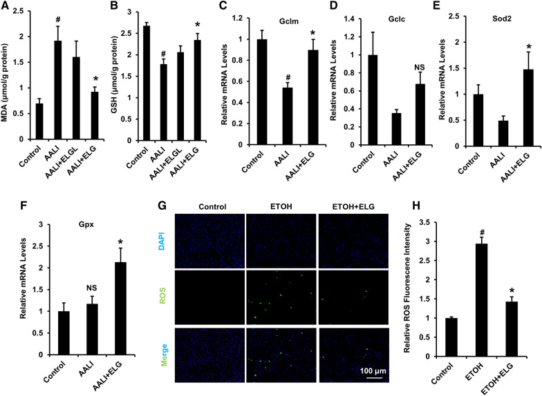
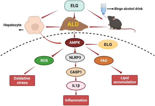
Background: LanGui tea, a traditional Chinese medicine formulation comprising of Gynostemma pentaphyllum (Thunb.) Makino, Cinnamomum cassia (L.) J. Presl, and Ampelopsis grossedentata (Hand-Mazz) W.T. Wang, has yet to have its potential contributions to alcoholic liver disease (ALD) fully elucidated. Consequently, the objective of this research is to investigate the protective properties of LanGui tea against binge alcohol-induced ALD and the mechanisms underlying its effects.

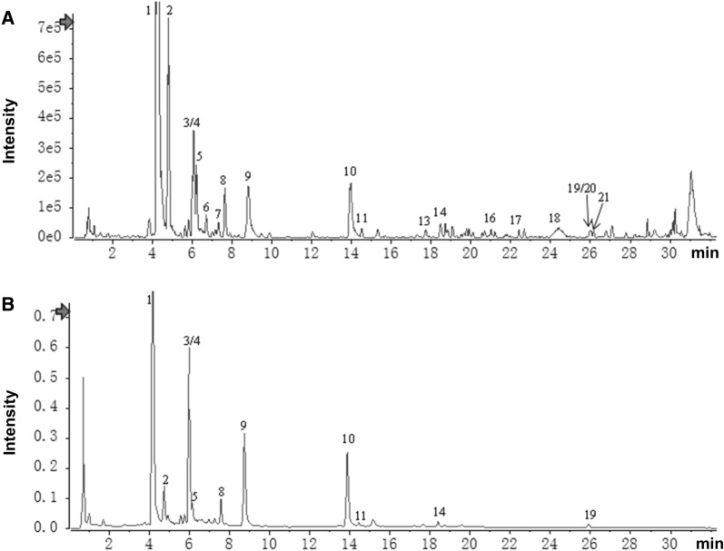
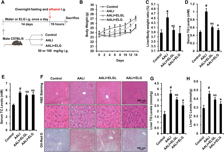
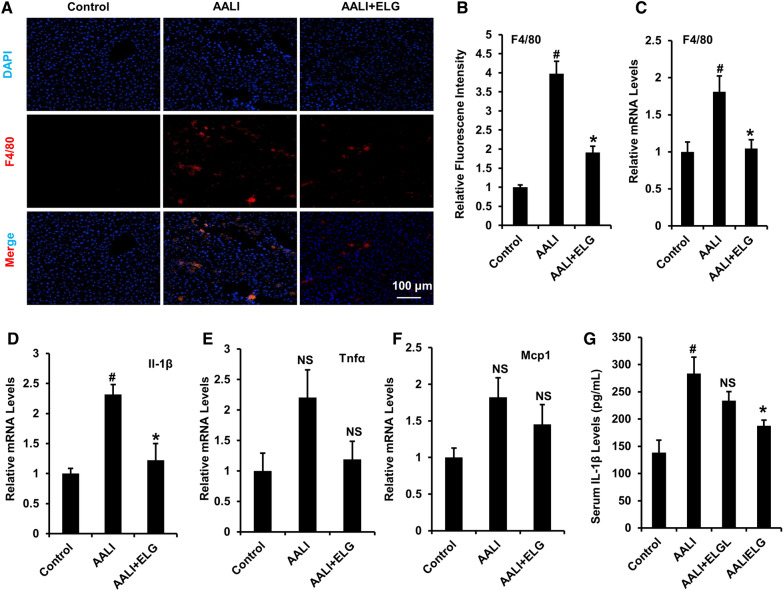
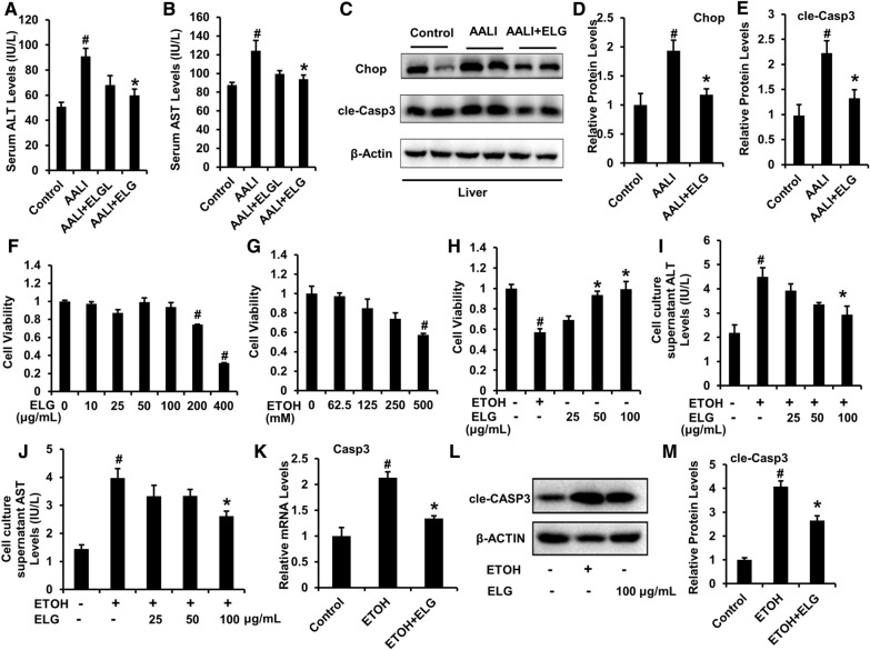
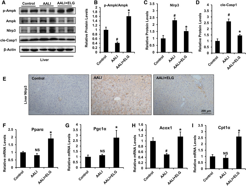
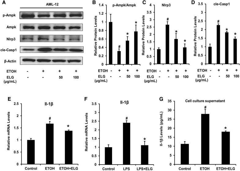
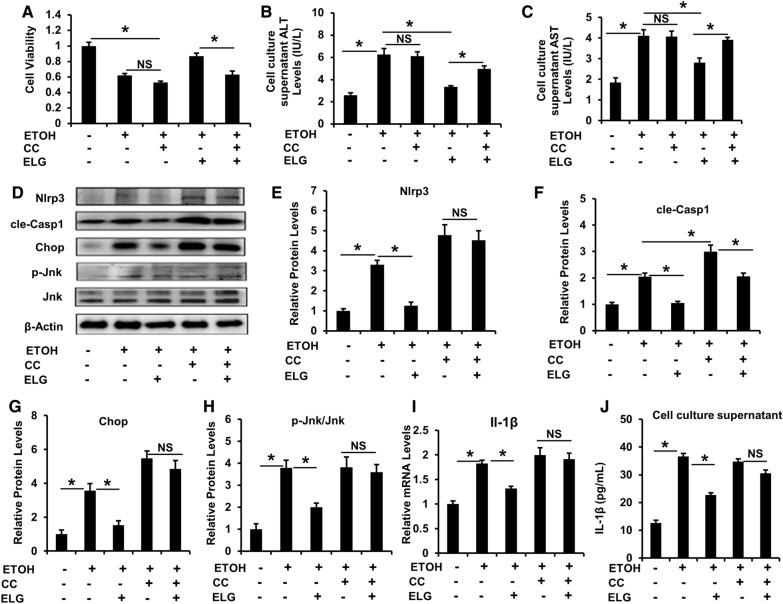
Methods: An experimental model of acute alcohol-induced liver disease was performed to assess the protective effects of extract of LanGui tea (ELG) at both 50 and 100 mg.kg-1 dosages on male C57BL/6 mice. Various parameters, including hepatic histological changes, inflammation, lipids content, as well as liver enzymes and interleukin 1β (IL-1β) in the serum were measured. The pharmacological mechanisms of ELG, specifically its effects on adenosine monophosphate-(AMP)-activated protein kinase (AMPK) and NLR family pyrin domain containing 3 (NLRP3) signaling, were investigated through Western blotting, qRT-PCR, ELISA, immunohistochemistry, immunofluorescence analyses, and by blocking the AMPK activity.

Results: ELG demonstrated a mitigating effect on fatty liver, inflammation, and hepatic dysfunction within the mouse model. This effect was achieved by activating AMPK signaling and inhibitingNLRP3 signaling in the liver, causing a reduction in IL-1β generation. In vitro studies further confirmed that ELG inhibited cell damage and IL-1β production in ethanol-induced hepatocytes by enhancing AMPK-NLRP3 signaling. Conversely, the pharmacological inhibition of AMPK activity nearly abrogated such alteration.

Conclusions: Thus, LanGui tea emerges as a promising herbal therapy for ALD management involving AMPK-NLRP3 signaling.

Keywords: Adenosine monophosphate-(AMP)-activated protein kinase; Alcoholic liver disease; Fatty liver; LanGui tea; NLR family pyrin domain containing 3.


Conflict of interest statement

No potential competing interests were reported.

Figures

Fig. 1
Fig. 1
Extract of LanGui tea (ELG) components analysis by Ultra-Performance Liquid Chromatography Quadrupole-Time of Flight Mass Spectrometer (UPLC-Q-TOF–MS). A Base peak ion (BPI) chromatogram of ELG in positive ion mode determined by UPLC-HRMS analysis; B UPLC-UV chromatogram of ELG (254 nm)
Fig. 2
Fig. 2
ELG combats acute alcohol-induced fatty liver in mice. A Overview of the animal experimental design. B Body weight curve of mice. n = 7. C Ratio of liver to body weight. n = 7, per group. D, E Serum TG and TC levels. n = 7. F Typical images of mouse liver tissues stained with H&E (original magnification, × 100; Scale bars, 200 µm) and oil red O (original magnification, × 200; Scale bars, 100 µm). n = 7. The white dotted rectangular frame indicates the area of inflammatory cell infiltration. G, H Mouse TG and TC levels in liver. n = 7. Statistical analysis of all experimental data was performed as means ± SEM. #p < 0.05, AALI vs. control group; *p < 0.05, AALI vs AALI + ELG group, NS not significance
Fig. 3
Fig. 3
ELG suppresses hepatic inflammation response in acute binge alcohol-treated mice. A, B Hepatic F4/80 immunofluorescence and relative fluorescence intensity analysis in mice. n = 5. Original magnification, × 200; Scale bars, 100 µm. CF Relative mRNA levels of hepatic pro-inflammatory cytokines. n = 5. G Serum Il-1β levels. n = 6. Statistical analysis of all experimental data was performed as means ± SEM. #p < 0.05, AALI vs. control group; *p < 0.05, AALI vs AALI + ELG group, NS not significance
Fig. 4
Fig. 4
ELG protects against acute ethanol (ETOH)—induced liver enzymes dysfunction and impairment of hepatocyte viability in mice and cell models. A, B Serum ALT and AST levels. n = 7. CE Mouse representative western blot images and relative protein levels in the liver. n = 5. #p < 0.05, AALI vs. control group; *p < 0.05, AALI vs AALI + ELG group. F, G CCK8 evaluation in AML-12 cells receiving 24-h DMSO, ELG (0, 10, 25, 50, 100, 200 and 400 µg.mL−1), or ethanol (0, 62.5, 125, 250 and 500 mM) treatment. n = 3. #p < 0.05, control vs ELG or ethanol group. AML-12 cells were incubated with DMSO and ELG (0, 25, 50 and 100 µg.mL−1) with or without ethanol (500 mM) for 24 h. H CCK8 assay. n = 3. I, J Cell culture supernatants ALT and AST levels. n = 4. (K) The relative mRNA level of Casp3 in AML-12 cells. n = 3. L, M Representative western blot image and quantitative data of protein expression of cle-Casp3 in AML-12 cells. n = 3. Experimental data were statistically analysed as the means ± SEM. #p < 0.05, ethanol vs. DMSO control group; *p < 0.05, ethanol vs ethanol + ELG group
Fig. 5
Fig. 5
ELG attenuates oxidative stress in liver and AML-12 cells under ethanol challenge. A, B Analysis of hepatic MDA and GSH contents. n = 6. CF mRNA determination of hepatic oxidative stress markers. n = 5. #p < 0.05, AALI vs. control group; *p < 0.05, AALI vs AALI + ELG group, NS not significance. G, H The representative fluorescent images and relative fluorescent intensity of ROS staining in AML-12 cells receiving 24-h co-incubation of ELG (100 µg.mL−1) and ethanol (500 mM). Original magnification, × 200; Scale bars, 100 µm. n = 3. All experimental data were statistically analysed as means ± SEM. #p < 0.05, ethanol vs. DMSO control group or *p < 0.05, ethanol vs ethanol + ELG group
Fig. 6
Fig. 6
ELG actives hepatic AMPK-NLRP3 signaling in acute binge alcohol-fed mice. Representative western blot image (A) and relative protein expression of p-Ampk/Ampk (B), Nlrp3/β-Actin (C), and cle-Casp1/β-Actin (D) in the liver of mice from control, AALI and AALI + ELG group. n = 5, per group. E Hepatic immunohistochemical analysis of Nlrp3. n = 5, per group. Original magnification, × 100; Scale bars, 200 µm. FI Relative mRNA levels of FAO-involved hepatic AMPK downstream genes. n = 5. Statistical analysis of all experimental data was performed as means ± SEM. #p < 0.05, AALI vs. control group; *p < 0.05, AALI vs AALI + ELG group, NS not significance
Fig. 7
Fig. 7
ELG regulates AMPK-NLRP3 signaling in mouse hepatocytes. AML-12 cells were treated by DMSO and ELG (0, 50 and 100 µg.mL−1) with or without ethanol (500 mM) for 24 h. Representative western blot image (A) and relative protein levels of genes (BD) in AMPK-NlRP3 signaling. n = 3, per group. E, F AML-12 cells receiving culture of DMSO, ethanol (500 mM) or LPS (100 ng.mL−1) and treated in the presence or absence of ELG (100 µg.mL−1) were used for subsequent analysis of relative mRNA levels of Il-1β. n = 3, per group. G Cell supernatant Il-1β concentration in AML-12 with ethanol (500 mM) stimulation and ELG (100 µg.mL−1) treatment. n = 3, per group. Data were statistically analysed as means ± SEM. #p < 0.05, as compared with control group; *p < 0.05, ELG treatment vs ethanol or LPS group
Fig. 8
Fig. 8
AMPK inactivation blunts the inhibitory effects of ELG on NLRP3 signaling and impairment in mouse hepatocytes. AML-12 cells insulted by ethanol (500 mM) were treated by DMSO and ELG (100 µg.mL−1) with or without CC (10 µM) for 24 h. A Cell viability assessment. n = 3. B, C ALT and AST levels in cell culture supernatants. n = 3. Representative western blot image (D) and relative protein expression of Nlrp3 (E), cle-Casp1 (F), Chop (G) and p-Jnk/Jnk (H) were displayed. n = 3. I Determination of relative mRNA expression of Il-1β. n = 3. J The cell supernatant Il-1β level. n = 4, per group. Data were statistically analysed as means ± SEM. *p < 0.05, as compared with ethanol group or ethanol + ELG + CC group, NS not significance
Fig. 9
Fig. 9
Proposed mechanisms of ELG in protecting acute binge alcohol-induced fatty liver. The main effect of ELG in protecting against acute alcohol-induced fatty liver may through activation of AMPK in hepatocytes, thus inhibiting NLRP3/Caspase1/Il-1β signaling. Consequently, ELG inhibits inflammation and oxidative stress injury in hepatocytes and promots hepatic fatty acid oxidation

References

    1. Seitz HK, Bataller R, Cortez-Pinto H, Gao B, Gual A, Lackner C, et al. Alcoholic liver disease. Nat Rev Dis Primers. 2018;4(1):16. doi: 10.1038/s41572-018-0014-7. - DOI - PubMed
    1. Singal AK, Mathurin P. Diagnosis and treatment of alcohol-associated liver disease: a review. JAMA. 2021;326(2):165–176. doi: 10.1001/jama.2021.7683. - DOI - PubMed
    1. Wu X, Fan X, Miyata T, Kim A, Cajigas-Du Ross CK, Ray S, et al. Recent advances in understanding of pathogenesis of alcohol-associated liver disease. Annu Rev Pathol. 2023;18:411–438. doi: 10.1146/annurev-pathmechdis-031521-030435. - DOI - PMC - PubMed
    1. Axley PD, Richardson CT, Singal AK. Epidemiology of alcohol consumption and societal burden of alcoholism and alcoholic liver disease. Clin Liver Dis. 2019;23(1):39–50. doi: 10.1016/j.cld.2018.09.011. - DOI - PubMed
    1. Barberia-Latasa M, Gea A, Martinez-Gonzalez MA. Alcohol, drinking pattern, and chronic disease. Nutrients. 2022;14(9):1954. doi: 10.3390/nu14091954. - DOI - PMC - PubMed

LinkOut - more resources