. 2024 Jun;11(21):e2306917.
doi: 10.1002/advs.202306917. Epub 2024 Mar 4.

Analyzing Mushroom Structural Patterns of a Highly Compressible and Expandable Hemostatic Foam for Gastric Perforation Repair

Affiliations

Analyzing Mushroom Structural Patterns of a Highly Compressible and Expandable Hemostatic Foam for Gastric Perforation Repair

Zhenzhen Shu et al. Adv Sci (Weinh). 2024 Jun.

Abstract

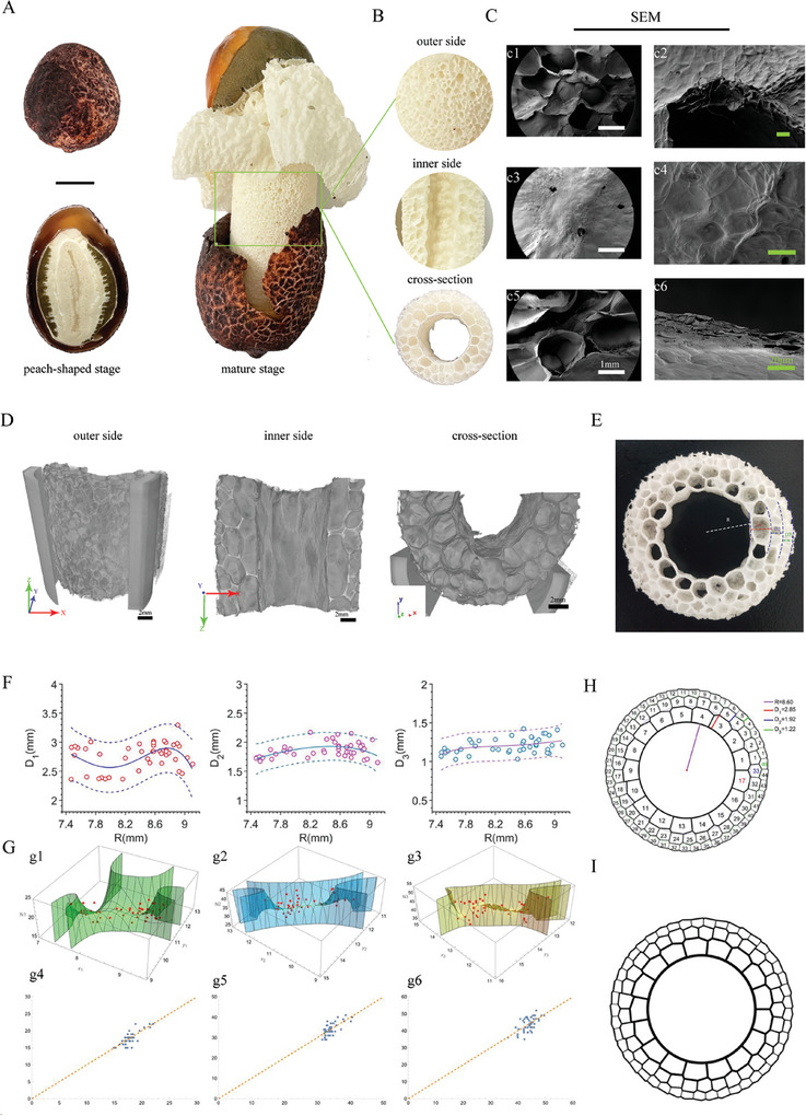
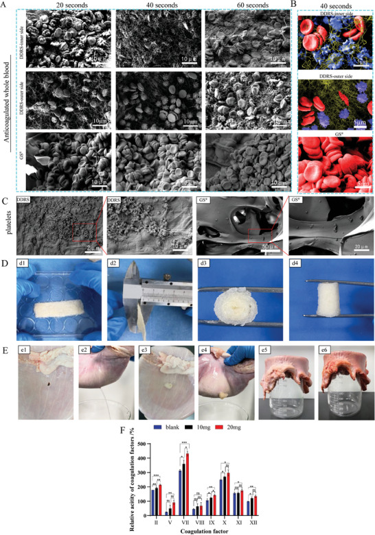
Nature presents the most beautiful patterns through evolving. Here, a layered porous pattern in golden ratio (0.618) is reported from a type of mushroom -Dictyophora Rubrovalvata stipe (DRS). The hierarchical structure shows a mathematical correlation with the golden ratio. This unique structure leads to superior mechanical properties. The gradient porous structure from outside to innermost endows it with asymmetrical hydrophilicity. A mathematical model is then developed to predict and apply to 3D printed structures. The mushroom is then explored to repair gastric perforation because the stomach is a continuous peristaltic organ, and the perforated site is subject to repeated mechanical movements and pressure changes. At present, endoscopic clipping is ineffective in treating ulcerative perforation with fragile surrounding tissues. Although endoscopic implant occlusion provides a new direction for the treatment of gastric ulcers, but the metal or plastic occluder needs to be removed, requiring a second intervention. Decellularized DRS (DDRS) is found with asymmetric water absorption rate, super-compressive elasticity, shape memory, and biocompatibility, making it a suitable occluder for the gastric perforation. The efficacy in blocking gastric perforation and promoting healing is confirmed by endoscopic observation and tissue analysis during a 2-month study.

Keywords: asymmetric hemostasis; gastric perforation; golden ratio in layered structure; gradient porous structure; mathematic modeling for 3D printing; super‐compressive elasticity.


Conflict of interest statement

The authors declare no conflict of interest.

Figures

Figure 1
Figure 1
Morphology and structure of DRS. A) The developmental process of Dictyophora rubrovalvata (DR). Left: peach‐shaped stage, right: mature stage. B) Enlarged view of the inner side, outer side and cross‐section of DRS. C) SEM images: (c1,c2) outer side, (c3,c4) inner side, (c5,c6) cross‐section at different magnifications. D) Micro‐CT images of the structure in different directions. E) Porous structure, R represents the cell radius, and D1, D2, viewand D3 represent the average inner diameter of each layer, respectively (Video S1, Supporting Information). F) Linear chart representing the relationship between R and D1, D2, and D3. G) Set the number of polygons in each layer to N1, N2, and N3, respectively. 2D curves representing the relationship between D, R and N (x1 = R, y1 = R+D1, x2 = R+D1, y2 = R+D1+D2, x3 = R+D1+D2, y3 = R+D1+D2+D3) (g1,g2,g3). Predictive calibration charts represent the relationship between the actual predicted value and the true value of N (g4,g5,g6). H,I) Computer simulated cross‐section image and the callout drawings of DRS.
Figure 2
Figure 2
3D simulated model. A) Simulated DRS model (a1), Biomimetic DRS model obtained by biological silicone printing (a2) (Video S2, Supporting Information), which can be folded (a3) and compressed (a4). B) A simulated DRS model. (b1) printed by biological silicone (2.5 cm in height, 8 mm in thickness), (b2) The stress–strain curve of the DRS model under cyclic compression in the direction parallel to the transverse plane, and images in compression experiments (Video S3, Supporting Information), (b3) The stress–strain curve of the DRS model under cyclic compression in the direction perpendicular to the transverse plane, and images in compression experiments. A concentric cylindrical model(Video S3, Supporting Information), (b4) printed by biological silicone (2.5 cm in height, 8 mm in thickness), (b5) The stress–strain curve of the concentric circle model under cyclic compression in the direction parallel to the transverse plane, and images in compression experiments, (b6) The stress–strain curve of the concentric circle model under cyclic compression in the direction perpendicular to the transverse plane, and images in compression experiments(n = 3, p <0.05).
Figure 3
Figure 3
Morphology and decellularization effect of DDRS. A) SEM images of the outer, inner, and cross‐section of DDRS. B) Images of different staining methods to observe the paraffin sections of DRS before and after acellularization (PAS, Giemsa, CWS). C) TEM images of the cross‐section of DDRS.
Figure 4
Figure 4
Water absorption of materials and different permeability of inner side and outer side of DDRS. A) Images on the inner and outer sides of DDRS before and after water absorption. B) Water absorption of DDRS at different time points. C) Permeability of inner and outer sides of DDRS at different times. The red liquid represents the diluted magenta solution (Video S5, Supporting Information). D) Permeability of inner and outer sides of DDRS at different times. The blue liquid represents diluted Giemsa solution, and the yellow liquid is diluted sodium fluorescein (Videos S6 and S7, Supporting Information). E) Blood permeability of inner and outer sides DDRS at different times (Videos S8–S10, Supporting Information).
Figure 5
Figure 5
Compression elasticity and shape memory of material. A) Schematic diagram of compression test direction. And images of cyclic compression at 90% strain of DDRS. B) Shape recovery ability of DDRS in water (Video S11, Supporting Information). C,E,G) Under the speed of 40 mm min−1, the compression curve of the GS in ten cycles, and the strain was 70%, 80%, and 90%, respectively. D,F,H) Under the speed of 40 mm min−1, the compression curve of DDRS was ten cycles, and the strain was 70%, 80%, and 90%, respectively (Video S12, Supporting Information).
Figure 6
Figure 6
Histocompatibility, and degradability of DDRS. A) Schematic diagram of subcutaneous implantation test of DDRS in rats (a1), and the operative photos of subcutaneous insertion (a2, a3) (n = 3). B) Pictures of subcutaneous masses after subcutaneous implantation for 2 weeks (b1, b4), 4 weeks (b2, b5), and 6 weeks (b3, b6). C) HE staining pictures at different time points. D) DMEM soaked with DDRS for 24 h in the extract (the concentration is 1, 0.75, 0.5, and 0.25 mgmL−1), after 1, 2, 3 days of culture, the cell viability was tested by CCK‐8(n = 5). E) the in vivo degradation curve (n = 3).
Figure 7
Figure 7
Hemostasis and repair of gastric perforation. A) SEM images of the inner and outer sides of DDRS after exposure to whole blood (sodium citrate) at different time points (20, 40, and 60 s). GS was used as the control group. B) SEM images after exposure to whole blood (sodium citrate) for 40 s. The blue area represents activated platelets, the red area is red blood cells, the yellow area represents the fibrin reticulum. C) SEM photos showing the adhesion of platelets on DDRS and GS. D) The preparation process of the DDRS occluder. Flatten the DDRS and then curl it along the long axis to get an onion roll‐like device (d1, d2). View pictures of the device from different perspectives (d3, d4). E) DDRS was used for sealing of a leaking in vitro pig stomach. A 5 mm wound was created on the pig stomach (e1), which caused a hole to leak (e2), DDRS was then employed to seal the gap(e3), following which the hole no longer leaked (e4). Put the pig stomach upon a beaker, pour the simulated gastric juice on the pig stomach, observe immediately (e5), and observe after 24 h(e6). F) The impact of DDRS on the activity of coagulation factors. (n = 3, mean ± SD, *** p <0.001, ** p <0.01, * p <0.05).
Figure 8
Figure 8
Evaluation of the closure and recovery of stomach perforations in pigs by endoscopic. A) Schematic diagram of treatment of gastric perforation pig. B) Create a perforation model of the stomach wall using an electric knife under endoscopic guidance. C) Put the material into the perforated part of the gastric wall (Video S13, Supporting Information). D) The material surface is sprayed with water to induce expansion, and the expanded material will seal the hole within 3 min. (Video S13, Supporting Information). E) Pictures of magnetic control bed (e1) and magnetic control capsule endoscopy (e2). F) Photos taken by magnetic capsule endoscopy after 7 days and 14 days of treatment. G) Specimens of perforated tissue of pig stomach. (g1) Mucosal side, (g2) Serous side. H) Pictures taken by endoscope 2 months after treatment and the healing and pathological results.

Similar articles

References

    1. Liu S., Luan Z., Wang T., Xu K., Luo Q., Ye S., Wang W., Dan R., Shu Z., Huang Y., ACS Nano. 2022, 17, 111. - PubMed
    1. Verlaan T., Voermans R. P., Henegouwen M. I. v. B., Bemelman W. A., Fockens P., Gastrointestinal Endoscopy. 2015, 82, 618. - PubMed
    1. Lanas A., García‐Rodríguez L. A., Polo‐Tomás M., Ponce M., Alonso‐Abreu I., Perez‐Aisa M. A., Perez‐Gisbert J., Bujanda L., Castro M., Muñoz M., Off. J. American College of Gastroenterology. 2009, 104, 1633. - PubMed
    1. Paspatis G. A., Dumonceau J. M., Barthet M., Meisner S., Repici A., Saunders B. P., Vezakis A., Gonzalez J. M., Turino S. Y., Tsiamoulos Z. P., Fockens P., Hassan C., Endoscopy. 2014, 46, 693. - PubMed
    1. Tarasconi A., Coccolini F., Biffl W., Tomasoni M., Ansaloni L., Picetti E., Molfino S., Shelat V., Cimbanassi S., Weber D., Abu‐Zidan F., Campanile F., Di Saverio S., Baiocchi G., Casella C., Kelly M., Kirkpatrick A., Leppaniemi A., Moore E., Peitzman A., Fraga G., Ceresoli M., Maier R., Wani I., Pattonieri V., Perrone G., Velmahos G., Sugrue M., Sartelli M., Kluger Y., et al., World journal of emergency surgery. 2020, 15, 3. - PMC - PubMed

LinkOut - more resources