Case Reports
. 2024 Feb 2;16(2):e53477.
doi: 10.7759/cureus.53477. eCollection 2024 Feb.

Cor Triatriatum Dexter With a Sinus Venosus Atrial Septal Defect in a 50-Year-Old Woman: A Case Report

Affiliations
Case Reports

Cor Triatriatum Dexter With a Sinus Venosus Atrial Septal Defect in a 50-Year-Old Woman: A Case Report

Hasan Kazma et al. Cureus. .

Abstract

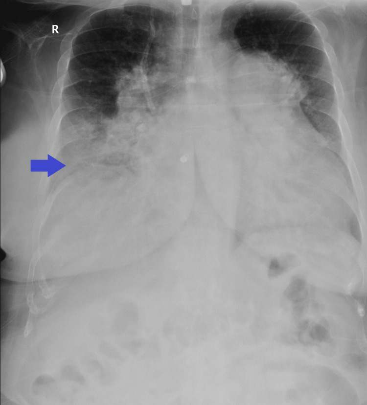
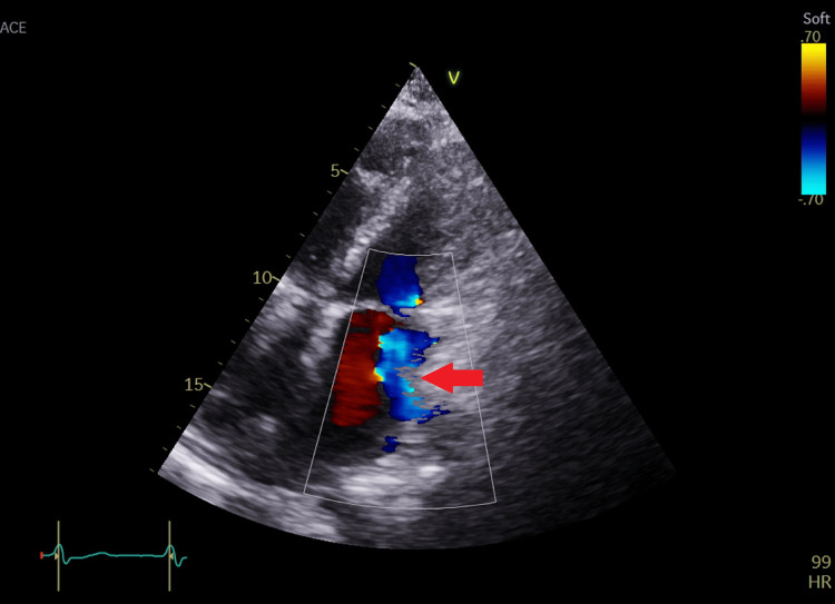
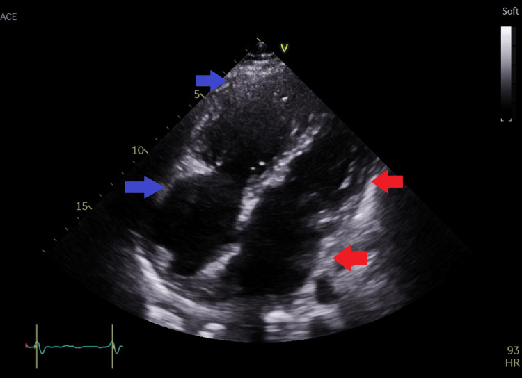
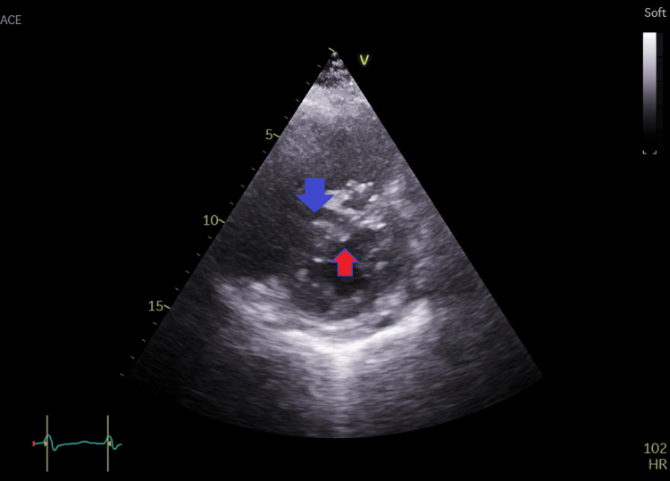
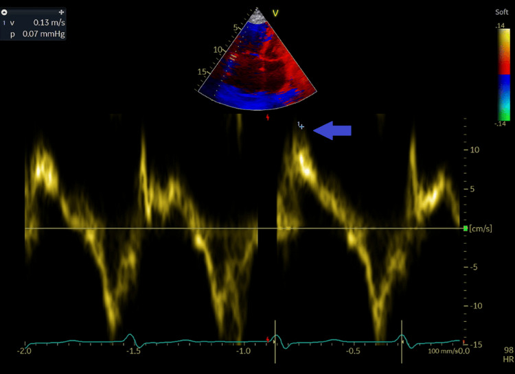
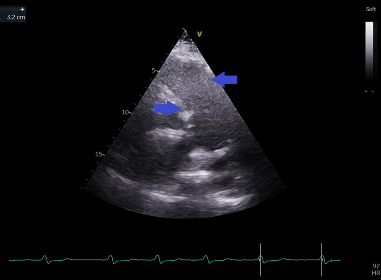
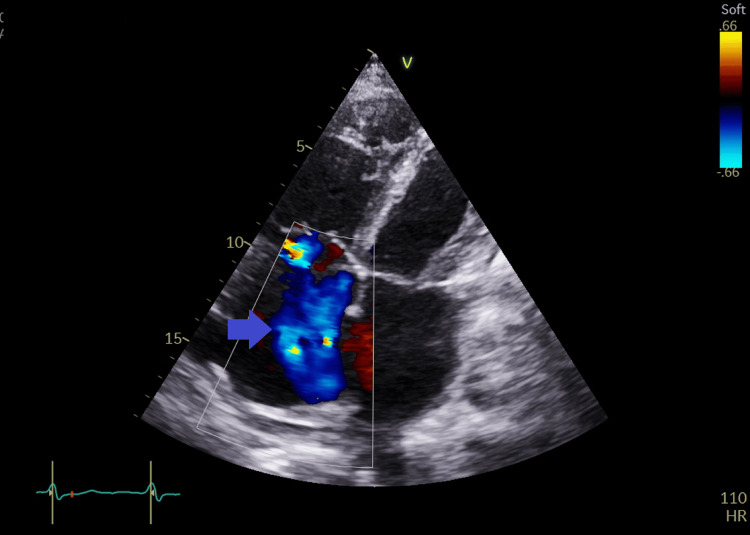
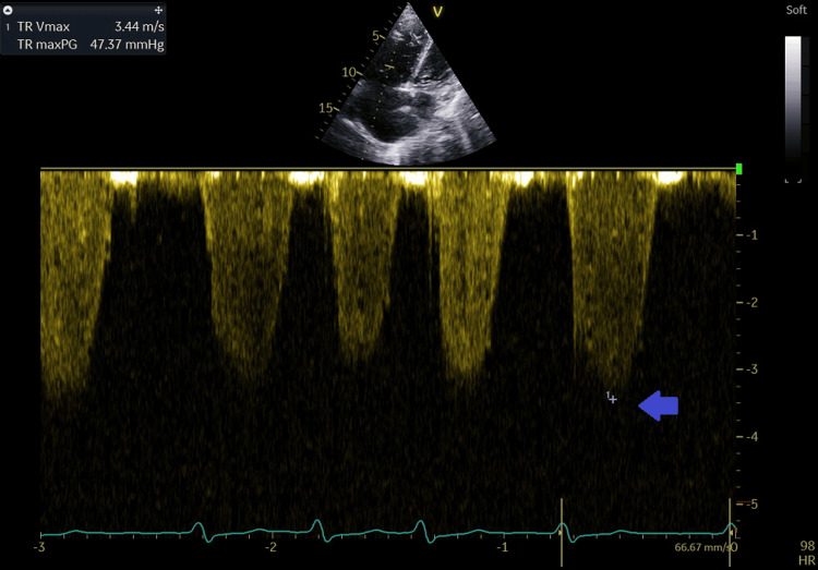
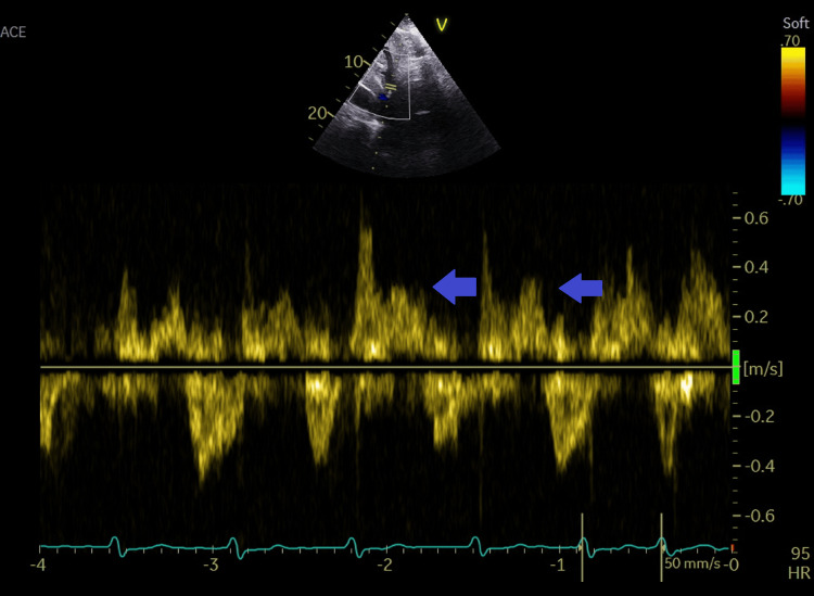
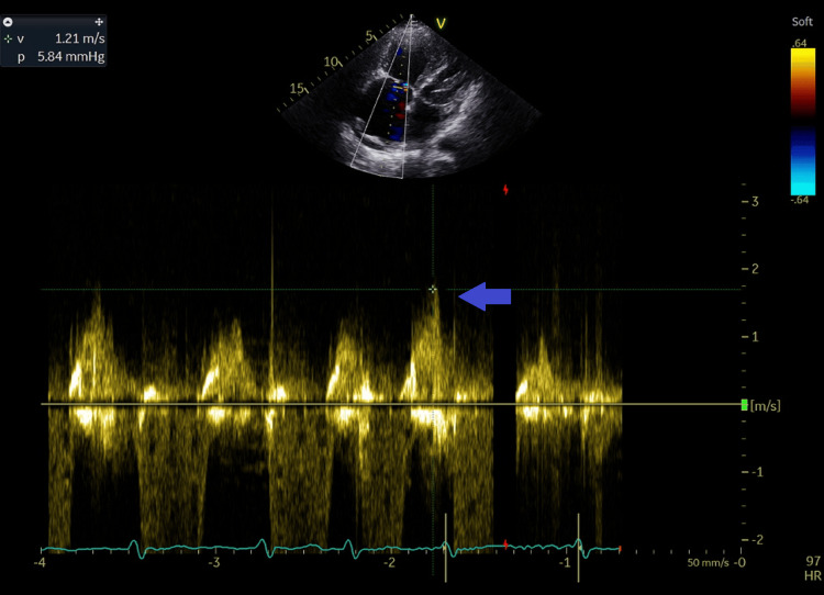
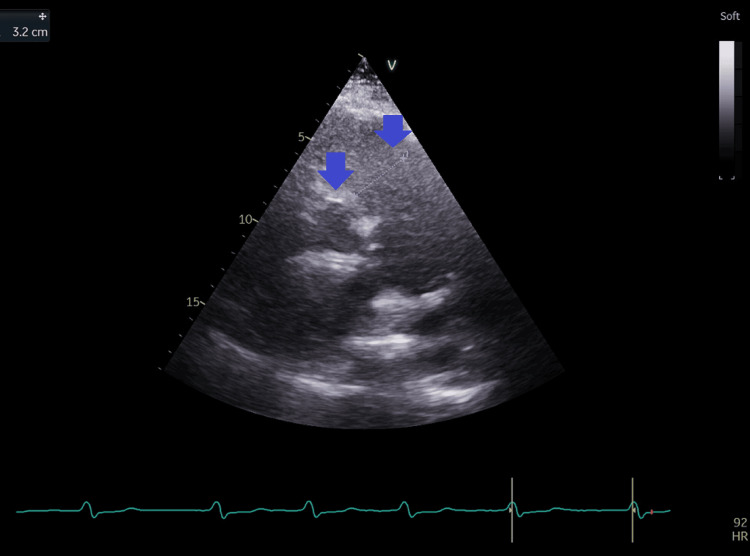
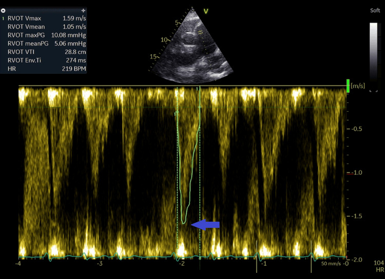
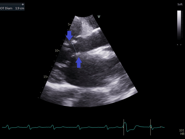
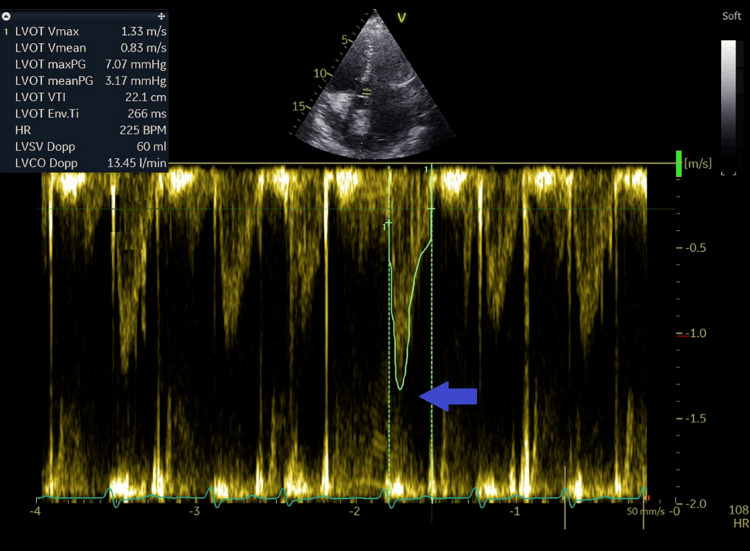
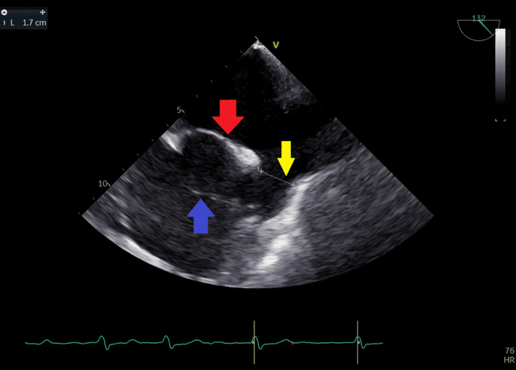
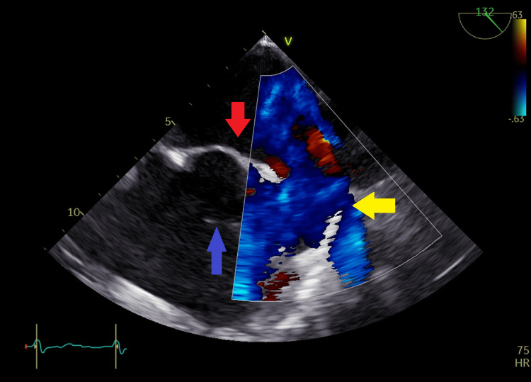
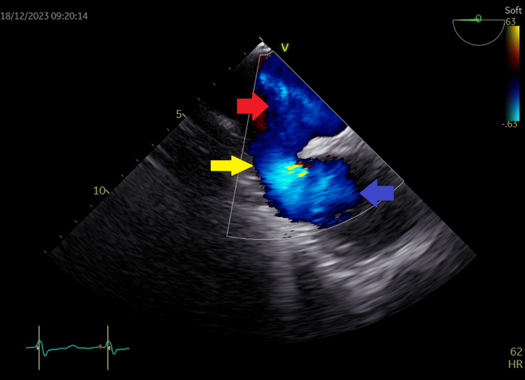
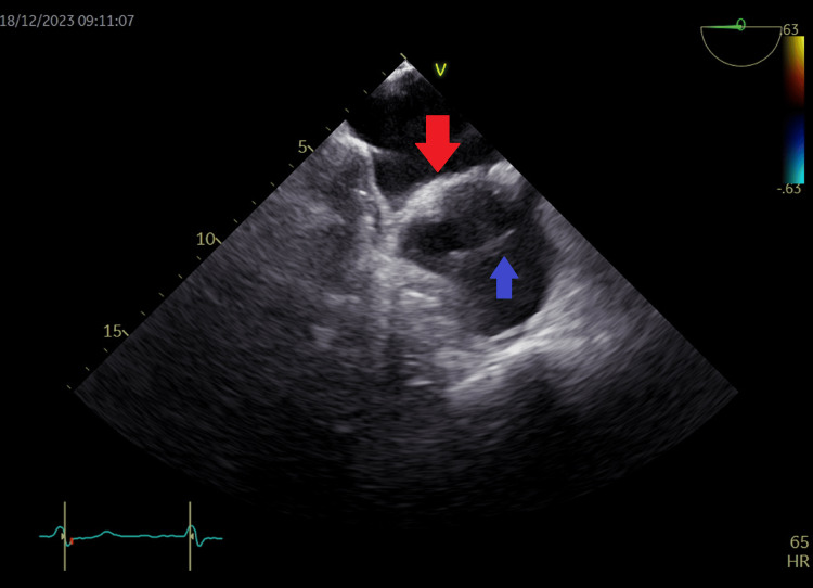
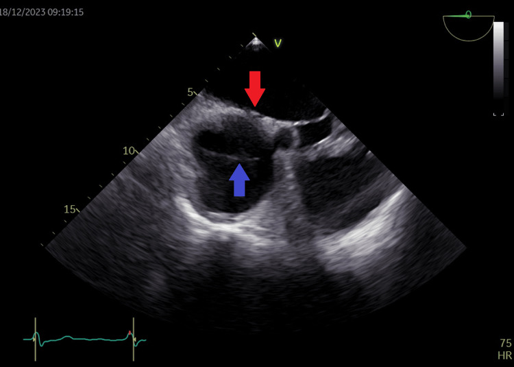
The diagnosis of atrial septal defect (ASD) may be delayed until adulthood or even later in life as it is a well-tolerated congenital heart disease. If patients are not examined and investigated well in childhood, the diagnosis may be delayed until later in adulthood when patients present with palpitations and sometimes dyspnea due to the right chambers dilatation from right ventricular volume overload. In this report, we present a case of a 50-year-old female patient with symptoms of heart failure and atrial fibrillation who was found to have dilated right cardiac chambers, dilated pulmonary artery, severe tricuspid regurgitation, pulmonary hypertension, and a pulmonary-to-systemic flow ratio (Qp/Qs) of more than 1.5 by transthoracic echocardiography and Doppler, indicating left to right shunt at the atrial level. However, transthoracic echocardiography could not visualize the defect, and two-dimensional (2D) transesophageal echocardiography was done in this patient and documented the presence of a sinus venosus ASD with an incomplete cor triatriatum dexter membrane; all four pulmonary veins were identified going to the left atrium. Since the presence of an incomplete cor triatriatum dexter membrane (despite causing no symptoms) makes the percutaneous closure of the sinus venosus ASD and the percutaneous repair of tricuspid regurgitation very difficult, we decided to advise surgical ASD closure and tricuspid valve repair for the patient.

Keywords: adult congenital heart disease; anomalous venous return; atrial septal defects; cor triatriatum dexter; functional tricuspid valve regurgitation; pulmonary hypertension; pulmonary to systemic flow ratio; right cardiac chambers dilatation; sinus venosus atrial septal defect; transthoracic and transesophageal echocardiography.


Conflict of interest statement

The authors have declared that no competing interests exist.

Figures

Figure 1
Figure 1. Chest X-ray showing cardiomegaly and right lower lobe infiltrates (blue arrow).
Figure 2
Figure 2. Apical four-chamber view with color coding of the mitral valve showing mild mitral regurgitation.
Figure 3
Figure 3. Modified apical four-chamber view showing dilated right atrium and right ventricle.
blue arrows=right atrium and right ventricle; red arrows=left atrium and left ventricle
Figure 4
Figure 4. Left parasternal short-axis view of the left and right ventricles showing dilated right ventricle (blue arrow) with right ventricular volume overload: D-shaped left ventricle (red arrow) in diastole
Figure 5
Figure 5. Tricuspid annulus velocity at lateral level by tissue pulsed wave Doppler from the apical window showing normal right ventricular systolic function with an S' at 13 cm/second (blue arrow).
Figure 6
Figure 6. Left parasternal short-axis view at the base of the heart showing a moderately dilated pulmonary artery (blue arrows).
Figure 7
Figure 7. Apical four-chambers view with color coding of the tricuspid valve showing severe tricuspid regurgitation (blue arrow).
Figure 8
Figure 8. Tricuspid regurgitation jet velocity recorded with continuous wave Doppler at 3.4 m/second (blue arrow).
Figure 9
Figure 9. Pulsed wave Doppler recording in the hepatic vein showing systolic reversal of flow, indicating severe tricuspid regurgitation.
Blue arrows show systolic reversal spectral display
Figure 10
Figure 10. Tricuspid E velocity by continuous wave Doppler recorded at 1.2 m/second from apical four-chamber view.
Blue arrow shows the E velocity spectral display
Figure 11
Figure 11. Short-axis view at the base of the heart from the left parasternal window with measurement of the right ventricular outflow tract diameter at 3.2 cm (blue arrows).
Figure 12
Figure 12. Short-axis view at the base of the heart from the left parasternal window with measurement of right ventricular outflow tract velocity at 1.59 m/second and time velocity integral at 28.8 cm (blue arrow).
Figure 13
Figure 13. Parasternal long-axis view with measurement of the left ventricular outflow tract diameter at 1.9 cm (blue arrow).
Figure 14
Figure 14. Measurement of the left ventricular outflow tract velocity at 1.3 m/seconds and time velocity integral at 21.1 cm from the apical five-chamber view (blue arrow).
Figure 15
Figure 15. Transesophageal echocardiography at the level of left atrium and right atrium showing a 1.7 cm sinus venosus defect (yellow arrow), a cor triatriatum dexter menbrane in the right atrium (blue arrow), and an intact osteum secundum septum (red arrow) in left atrium.
Figure 16
Figure 16. Transesophageal echocardiography at the level of left atrium and right atrium with color flow coding at the sinus venosus site showing left to right shunt across the sinus venosus atrial septal defect.
Yellow arrow shows color-coded left-to-right shunt; blue arrow in right atrium shows part of the cor triatriatum dexter membrane; red arrow in left atrium shows intact osteum secundum septum
Figure 17
Figure 17. Transesophageal echocardiogram at the level of left atrium and right atrium showing color flow across the sinus venosus atrial septal defect with left to right shunt (yellow arrow). The red arrow shows flow from the left atrium to the right atrium (blue arrow).
Figure 18
Figure 18. Transesophageal echocardiography view of the left atrium (red arrow) and right atrium (blue arrow) also showing the cor triatriatum dexter membrane dividing the right atrium (blue arrow).
Figure 19
Figure 19. Transesophageal echocardiography view of the left atrium (red arrow) and right atrium (blue arrow) showing the cor triatriatum dexter membrane dividing the right atrium (blue arrow).

References

    1. Atrial septal defect in adulthood: a new paradigm for congenital heart disease. Brida M, Chessa M, Celermajer D, et al. Eur Heart J. 2022;43:2660–2671. - PubMed
    1. Correlation of transthoracic echocardiography-derived pulmonary to systemic flow ratio with hemodynamically estimated left to right shunt in atrial septal defects. Faherty E, Rajagopal H, Lee S, Love B, Srivastava S, Parness IA, Uppu SC. Ann Pediatr Cardiol. 2022;15:20–26. - PMC - PubMed
    1. Diagnosis of sinus venosus atrial septal defect with transesophageal echocardiography. Pascoe RD, Oh JK, Warnes CA, Danielson GK, Tajik AJ, Seward JB. Circulation. 1996;94:1049–1055. - PubMed
    1. Partial anomalous pulmonary venous connection: a great imitator? Seecheran RV, Dookie T, Seecheran VK, et al. J Investig Med High Impact Case Rep. 2020;8:2324709620933425. - PMC - PubMed
    1. Sinus venosus atrial septal defect: a challenging diagnosis. Vodusek Z, Khaliqdina S, Borz-Baba C, Scandrett R. Cureus. 2019;11:0. - PMC - PubMed

Publication types

LinkOut - more resources