. 2024 Feb 28:36:62-82.
doi: 10.1016/j.bioactmat.2024.02.024. eCollection 2024 Jun.

Controlled-release hydrogel loaded with magnesium-based nanoflowers synergize immunomodulation and cartilage regeneration in tendon-bone healing

Affiliations

Controlled-release hydrogel loaded with magnesium-based nanoflowers synergize immunomodulation and cartilage regeneration in tendon-bone healing

Jintao Li et al. Bioact Mater. .

Abstract

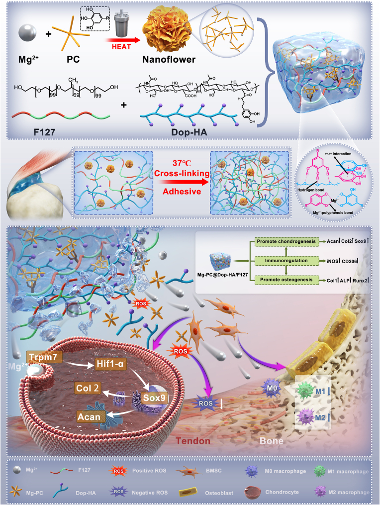
Tendon-bone interface injuries pose a significant challenge in tissue regeneration, necessitating innovative approaches. Hydrogels with integrated supportive features and controlled release of therapeutic agents have emerged as promising candidates for the treatment of such injuries. In this study, we aimed to develop a temperature-sensitive composite hydrogel capable of providing sustained release of magnesium ions (Mg2+). We synthesized magnesium-Procyanidin coordinated metal polyphenol nanoparticles (Mg-PC) through a self-assembly process and integrated them into a two-component hydrogel. The hydrogel was composed of dopamine-modified hyaluronic acid (Dop-HA) and F127. To ensure controlled release and mitigate the "burst release" effect of Mg2+, we covalently crosslinked the Mg-PC nanoparticles through coordination bonds with the catechol moiety within the hydrogel. This crosslinking strategy extended the release window of Mg2+ concentrations for up to 56 days. The resulting hydrogel (Mg-PC@Dop-HA/F127) exhibited favorable properties, including injectability, thermosensitivity and shape adaptability, making it suitable for injection and adaptation to irregularly shaped supraspinatus implantation sites. Furthermore, the hydrogel sustained the release of Mg2+ and Procyanidins, which attracted mesenchymal stem and progenitor cells, alleviated inflammation, and promoted macrophage polarization towards the M2 phenotype. Additionally, it enhanced collagen synthesis and mineralization, facilitating the repair of the tendon-bone interface. By incorporating multilevel metal phenolic networks (MPN) to control ion release, these hybridized hydrogels can be customized for various biomedical applications.

Keywords: Controlled release; Immunomodulation; Magnesium; Metal–phenolic networks; Self-assembly process; Tendon-bone interface.


Conflict of interest statement

The authors declared that they have no conflicts of interest to this work. We declare that we do not have any commercial or associative interest that represents a conflict of interest in connection with the work submitted.

Figures

Image 1
Graphical abstract
Fig. 1
Fig. 1
Schematic synthesis and characterization of Mg-PC. A) Schematic synthesis of Mg-PC by hydrothermal method; B) XRD patterns of PC and Mg-PC; C) FTIR spectra of MgCl2, PC, and Mg-PC; D) TGA curves of Mg-PC; E) Scanning electron microscope images and magnified images of Mg-PC; F) Particle size distribution of Mg-PC; G) Elemental mapping of Mg-PC.
Scheme 1
Scheme 1
Schematic representation of the design of an injectable composite hydrogel. This hydrogel could control the release of Mg-PC for bone-tendon interface repair by modulating the inflammatory environment and promoting cartilage regeneration.
Fig. 2
Fig. 2
Mechanism and characterization of injectable composite hydrogels. A) Schematic diagram of the mechanism of injectable composite hydrogels; B) Gel characteristics (temperature and time) of composite hydrogels with different contents of Mg-PC (0%, 5%, 10%, and 20%); C) FTIR spectra of Mg-PC, Dop-HA, F127, and 10% Mg– PC@Dop-HA/F127; D) Macroscopic photos of hydrogels at different temperatures (Scale bars indicate 1 cm); E) Injectability, thermosensitivity and shape adaptability; F) Scanning electron microscopy images of composite hydrogels with different contents of Mg-PC (0%, 5%, 10%, and 20%); G) The elastic modulus (G′) and viscosity modulus (G″), and the rheological recovery behavior (H) of composite hydrogels with different contents of Mg-PC (0%, 5%, 10% and 20%); I) Mechanical properties of composite hydrogels with different contents of Mg-PC (0%, 5%, 10% and 20%); J) Weight loss of composite hydrogels with different contents of Mg-PC (0%, 5%, 10% and 20%); and release characteristics of Mg2+ (K) and PC (L) with different contents of Mg-PC (5%, 10% and 20%).
Fig. 3
Fig. 3
In vitro cytocompatibility and cell migration properties of composite hydrogels. A) Cytotoxicity of degradation products of composite hydrogels with different contents of Mg-PC (0%, 5%, 10% and 20%) on BMSCs; B) Live/dead images of BMSCs treated with 100-fold degradation products of different composite hydrogels for 24 h; C) Schematic diagram of the Transwell assay; D) Transwell images and migration rates of different composite hydrogels quantification (F); E) Cell migration images and related quantitative data obtained from scratch experiments (G); H) and I) are qRT-PCR results of CCL2 and CCL3 mRNA expression of BMSCs 3 days after treatment with degradation products of different hydrogels. *p < 0.05, **p < 0.01, “ns” represents no significant difference.
Fig. 4
Fig. 4
Antioxidant activity of Mg-PC and Mg-PC@Dop-HA/F127. A) Schematic diagram of the DPPH radical reduction reaction; B) Photographs of DPPH solution, MgCl2-treated DPPH solution, and Mg-PC-treated DPPH solution at different times; C) UV–visible spectra of the different modes of treatment and D) DPPH scavenging rate (n = 3); E) UV–visible spectral changes of DPPH solutions treated by Mg-PC for different times; F) UV–vis curves of Mg-PC; H) Fluorescence images and G) relative fluorescence intensities of oxidation inhibition obtained by ROS test kit on BMSCs (n = 3). *p < 0.05, **p < 0.01, ***p < 0.001,"ns” represents no significant difference.
Fig. 5
Fig. 5
Immunomodulatory activity of composite hydrogels. A) Schematic diagram of the experiment to study the polarizing effect of hydrogel on M0 macrophages; B) and C) Immunofluorescence staining of iNOS and CD206 after induction of M0 macrophages by hydrogel degradation products; D) and E) Quantification of immunofluorescence staining of iNOS and CD206; F) Western blotting showing expression of iNOS,CD206 and GAPDH. G) Representative images of RAW264.7 surface markers (CD86 and CD206) analyzed by flow cytometry. H) RT-qPCR measurements of gene expression of iNOS, TNF-α, CD206, and Arg after induction of M0 macrophages by hydrogel degradation products for 3 days. *p < 0.05, **p < 0.01, ***p < 0.001.
Fig. 6
Fig. 6
Osteogenic differentiation-promoting activity of composite hydrogels and mechanism of promoting chondrogenic differentiation. A) Alkaline phosphatase (ALP) staining images and alizarin red staining images; B) Quantification of ALP activity (at day 14); C) Alizarin red staining of the corresponding quantified red areas (at day 21); D) Western blotting bands of Col I, ALP and Runx2 after hydrogel degradation products in osteogenic medium treated BMSCs; (E) Toluidine blue staining images of hydrogel degradation products after treatment of BMSC pellets in osteogenic medium; (F) Western blotting bands of Acan, Col II and Sox9 after hydrogel degradation products in chondrogenic medium treated BMSCs; G) and H) RT-qPCR measurements of Sox9 and Col II gene expression by hydrogel degradation products after treating BMSCs in chondrogenic medium for 7 days; I) Toluidine blue staining images of hydrogel degradation products and Mg2+ channel inhibitor (2APB) after chondrogenic medium treatment of BMSC pellets; J) Images of immunofluorescence staining (Col II and Aggrecan) and corresponding fluorescence intensity quantification (N) of hydrogel degradation products and Mg2+ channel inhibitor (2APB) in chondrogenic medium after treatment of BMSC pellets; K) Representative images of Mg2+ analyzed by flow cytometry; L) Immunofluorescence photos of Mg2+ labeling and associated quantification (M); O) Western blot bands of TRPM7, Hif-1α, Acan, Col II, and Sox9 by hydrogel degradation products and Mg2+ channel inhibitor (2APB) after treating BMSCs in chondrogenic medium for 7 days.(P) RT-qPCR measurements of Sox9, Col II and Aggrecan gene expression by hydrogel degradation products and Mg2+ channel inhibitor (2APB) in chondrogenic medium after treating BMSCs for 7 days.*p < 0.05, **p < 0.01, ***p < 0.001,"ns” represents no significant difference.
Fig. 7
Fig. 7
Micro-CT analysis results and Data for biomechanical tests of in vivo animal experiments. A) Schematic of the animal experiments; B) Three-dimensional reconstruction images and coronal images of the proximal humerus micro CT of rats at 4 and 8 weeks after surgery; C) Images of a finite element model of the proximal humerus in rats at 4 and 8 weeks after surgery; D) Images of biomechanical tests; E) BMD,BV/TV and Tb. Sp values of the tendon-to-bone interface; F) Data for biomechanical tests, including maximum load, stiffness, and distance at maximum load.*p < 0.05, **p < 0.01, “ns” represents no significant difference.
Fig. 8
Fig. 8
Morphological analysis of newly formed tendon-bone interface tissues after treatment. A) Representative images of hematoxylin and eosin staining (H&E) and toluidine blue/fast green staining (B) of slices in rats treated with normal, suture, Dop-HA/F127, and Mg-PC@Dop-HA/F127 groups, respectively. T: tendon; I: Interface; B: Bone. C) Sirius red stained photos of different treatment groups; D) Bone-tendon interface maturing score of different treatment groups (n = 3) and E) the area of newly formed fibrocartilage in the different treatment groups (n = 3). Scale bars indicate 200 μm for 40 × and 100 μm for 100 × .*p < 0.05, **p < 0.01.
Fig. 9
Fig. 9
Immunohistochemical staining of newly formed tendon bone interface tissues after treatment. A) Immunohistochemical staining images and associated expression levels of Trpm7 and Hif-1α at the tendon bone interface in different treatment groups (n = 3) at 4 and 8 weeks after treatment (B&C). D) Immunohistochemical staining images and associated expression levels of Col II and Sox9 at the tendon bone interface in different treatment groups (n = 3) at 4 and 8 weeks after treatment (E&F). Scale bars indicate 200 μm for 40 × and 100 μm for 100 × .*p < 0.05, **p < 0.01.
Fig. 10
Fig. 10
Immunofluorescence (CD44 and CD206) staining results. A) Immunofluorescence and relative intensity quantification(C) of CD44 at the tendon bone interface at 4 and 8 weeks after treatment in different groups; B) Immunofluorescence and relative intensity quantification(D) of CD206 at the tendon bone interface at 4 weeks after treatment in different groups. **p < 0.01, ***p < 0.001,"ns” represents no significant difference.

Similar articles

Cited by

References

    1. Font Tellado S., Balmayor E.R., Van Griensven M. Strategies to engineer tendon/ligament-to-bone interface: biomaterials, cells and growth factors. Adv. Drug Deliv. Rev. 2015;94:126–140. doi: 10.1016/j.addr.2015.03.004. - DOI - PubMed
    1. Shaw H.M., Benjamin M. Structure-function relationships of entheses in relation to mechanical load and exercise. Scand. J. Med. Sci. Sports. 2007;17(4):303–315. doi: 10.1111/j.1600-0838.2007.00689.x. - DOI - PubMed
    1. Atesok K., Doral M.N., Karlsson J., Egol K.A., Jazrawi L.M., Coelho P.G., Martinez A., Matsumoto T., Owens B.D., Ochi M., Hurwitz S.R., Atala A., Fu F.H., Lu H.H., Rodeo S.A. Multilayer scaffolds in orthopaedic tissue engineering. Knee Surg. Sports Traumatol. Arthrosc. J. ESSKA. 2016;24(7):2365–2373. doi: 10.1007/s00167-014-3453-z. - DOI - PubMed
    1. Lim W.L., Liau L.L., Ng M.H., Chowdhury S.R., Law J.X. Current progress in tendon and ligament tissue engineering. Tissue Eng. Regen. Med. 2019;16(6):549–571. doi: 10.1007/s13770-019-00196-w. - DOI - PMC - PubMed
    1. Wang D., Zhang X., Huang S., Liu Y., Fu B.S., Mak K.K., Blocki A.M., Yung P.S., Tuan R.S., Ker D.F.E. Engineering multi-tissue units for regenerative Medicine: bone-tendon-muscle units of the rotator cuff. Biomaterials. 2021;272 doi: 10.1016/j.biomaterials.2021.120789. - DOI - PubMed

LinkOut - more resources