

The Rat Thoracic Ultrasound protocol: scanning technique and normal findings
- PMID: 38440385
- PMCID: PMC10909930
- DOI: 10.3389/fvets.2024.1286614
The Rat Thoracic Ultrasound protocol: scanning technique and normal findings
Erratum in
-
Erratum: The Rat Thoracic Ultrasound protocol: scanning technique and normal findings.Front Vet Sci. 2024 Apr 11;11:1408432. doi: 10.3389/fvets.2024.1408432. eCollection 2024. Front Vet Sci. 2024. PMID: 38665770 Free PMC article.
Abstract
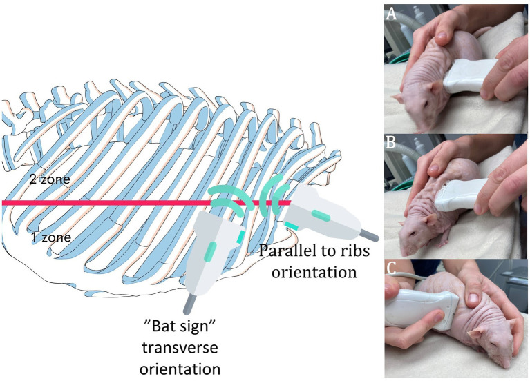
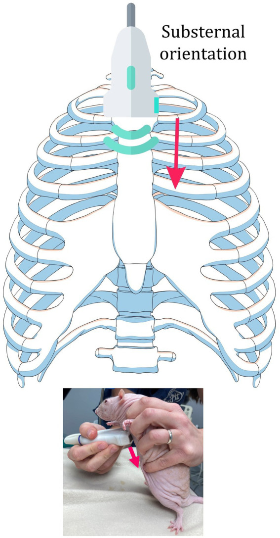
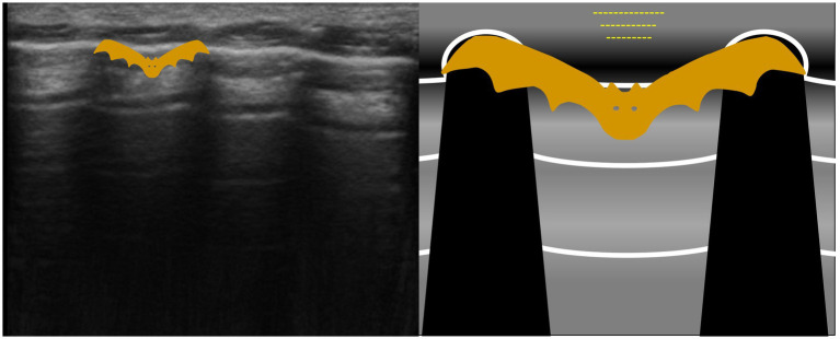
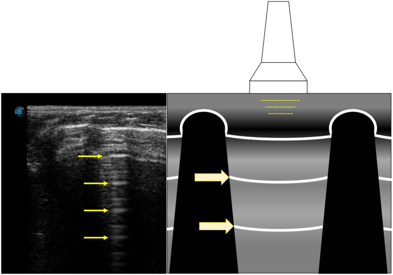
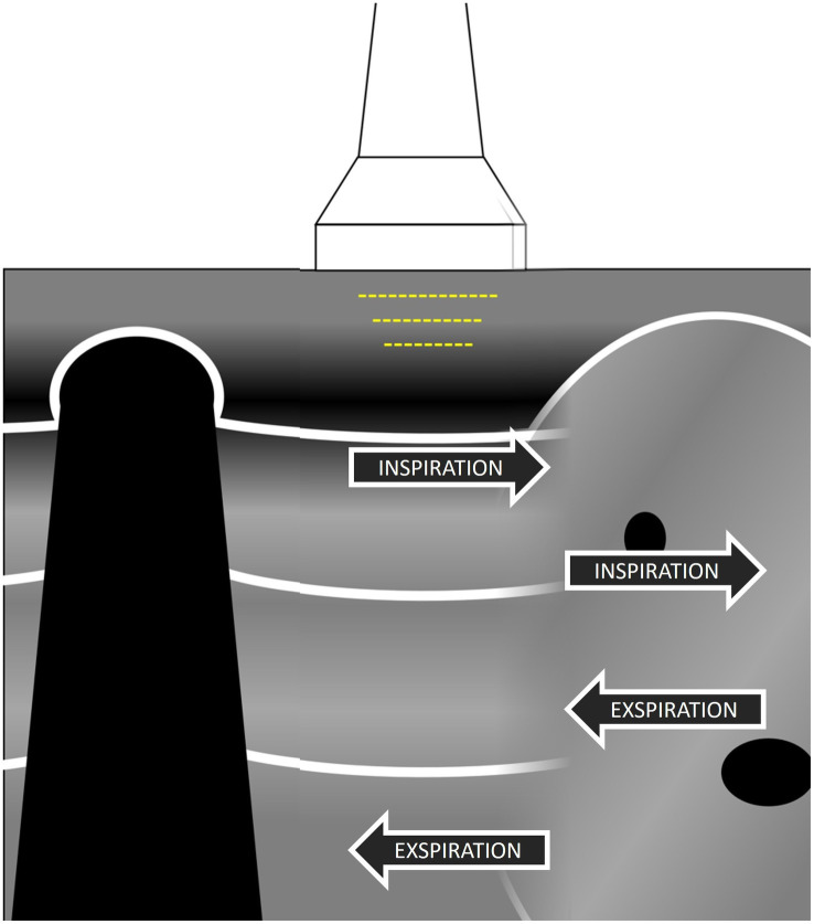
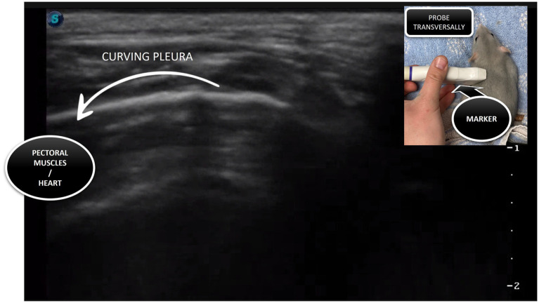
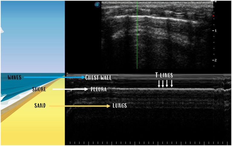
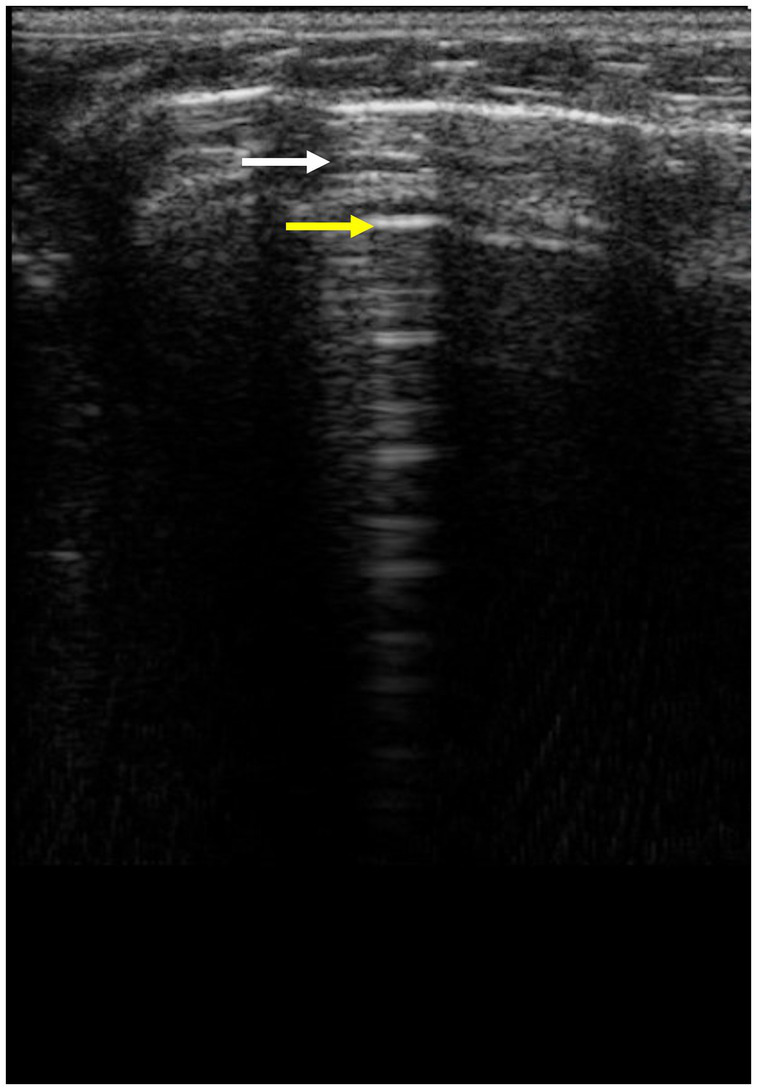
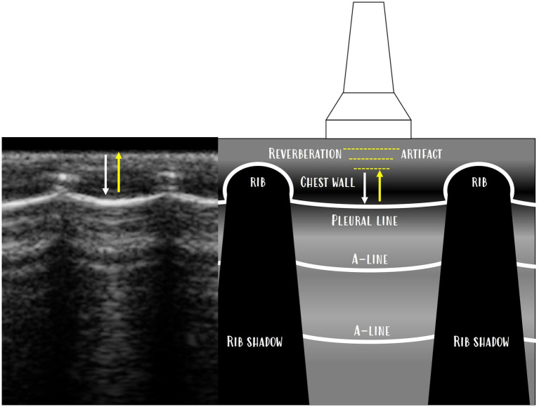
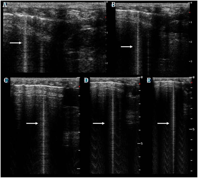
Respiratory diseases (especially pneumonia) are very common disorders in pet rats. The suspected diagnosis is mostly based on the clinical signs, thoracic auscultation, and thoracic radiography. However, auscultation is insensitive in determining the severity of the disease, and radiographs are often unremarkable. Non-cardiac thoracic ultrasonography is increasingly used in veterinary medicine; however, it has not been described in detail in rats. Thoracic ultrasonic examination was conducted on 400 client-owned conscious pet rats. The rats were examined in the period from June 2023 to August 2023 in two veterinary clinics. Due to the small size of the animal, different anatomical considerations, and different evaluation protocols, as well as to meet the optimal outcome of detailed thoracic ultrasound, a standard methodological protocol was developed, and the name RATTUS (Rat Thoracic Ultrasound) was proposed. Typical signs of normal RATTUS were described (bat sign, lung sliding, A-lines, abdominal curtain sign, ski jump sign, lung pulse, seashore sign in M-mode, and bamboo sign). The new evaluation of lung inflation symmetry by substernal access was also described. The methodical approach presented and the normal findings description are proposed to be used for a standard/routine thoracic ultrasound examination in pet rats.
Keywords: RATTUS; lung; pleura; rat; respiratory disease; thorax; ultrasonography.
Copyright © 2024 Piskovská, Kraszewska, Hauptman and Jekl.
Conflict of interest statement
The authors declare that the research was conducted in the absence of any commercial or financial relationships that could be construed as a potential conflict of interest.
Figures
Similar articles
-
RATTUS (Rat Thoracic Ultrasound): diagnosis of pneumothorax in pet rats.Front Vet Sci. 2024 Sep 13;11:1394291. doi: 10.3389/fvets.2024.1394291. eCollection 2024. Front Vet Sci. 2024. PMID: 39346960 Free PMC article.
-
Zombie Cruise Ship Virtual Escape Room for POCUS Pulmonary: Scan Your Way Out.J Educ Teach Emerg Med. 2022 Jul 15;7(3):SG1-SG23. doi: 10.21980/J8RM0M. eCollection 2022 Jul. J Educ Teach Emerg Med. 2022. PMID: 37465772 Free PMC article.
-
Abnormal Curtain Signs Identified With a Novel Lung Ultrasound Protocol in Six Dogs With Pneumothorax.Front Vet Sci. 2019 Aug 28;6:291. doi: 10.3389/fvets.2019.00291. eCollection 2019. Front Vet Sci. 2019. PMID: 31555674 Free PMC article.
-
Ten good reasons to practice ultrasound in critical care.Anaesthesiol Intensive Ther. 2014 Nov-Dec;46(5):323-35. doi: 10.5603/AIT.2014.0056. Anaesthesiol Intensive Ther. 2014. PMID: 25432552 Review.
-
[Lung ultrasound in acute and critical care medicine].Anaesthesist. 2012 Jul;61(7):608-17. doi: 10.1007/s00101-012-2046-9. Anaesthesist. 2012. PMID: 22772347 Review. German.
Cited by
-
RATTUS (Rat Thoracic Ultrasound): diagnosis of pneumothorax in pet rats.Front Vet Sci. 2024 Sep 13;11:1394291. doi: 10.3389/fvets.2024.1394291. eCollection 2024. Front Vet Sci. 2024. PMID: 39346960 Free PMC article.
References
-
- Seiler G. Thorax In: Barr F Jd f, Gaschen L, editors. BSAVA manual of canine and feline ultrasonography. Great Britain: BSAVA; (2011). 29–36.
-
- Boysen S, Gommeren K, Chalhoub S. The essentials of veterinary point of care ultrasound: Pleural space and lung. Spain: Edra; (2022). 192 p.
LinkOut - more resources
Full Text Sources