

Evaluation of Anatomical Variations Associated with Chronic Rhinosinusitis by Computed Tomography of Paranasal Sinuses
- PMID: 38440475
- PMCID: PMC10908970
- DOI: 10.1007/s12070-023-04320-0
Evaluation of Anatomical Variations Associated with Chronic Rhinosinusitis by Computed Tomography of Paranasal Sinuses
Abstract
Background: Chronic rhinosinusitis (CRS) is a syndrome with multifactorial aetiology. Amongst which, anatomical variations studied by computed tomography of paranasal sinuses (CT PNS) had a high incidence which varied between 64.0% and 99.8%10. Due to such high incidence, this study is undertaken to assess the various anatomical variations and their significant association in CRS.
Method: A prospective observational study was conducted in 70 CRS patients and were subjected to CT PNS. CT PNS is studied to know the various anatomical variations & other CT findings causing CRS and then findings noted down, tabulated and statistical analysis done.
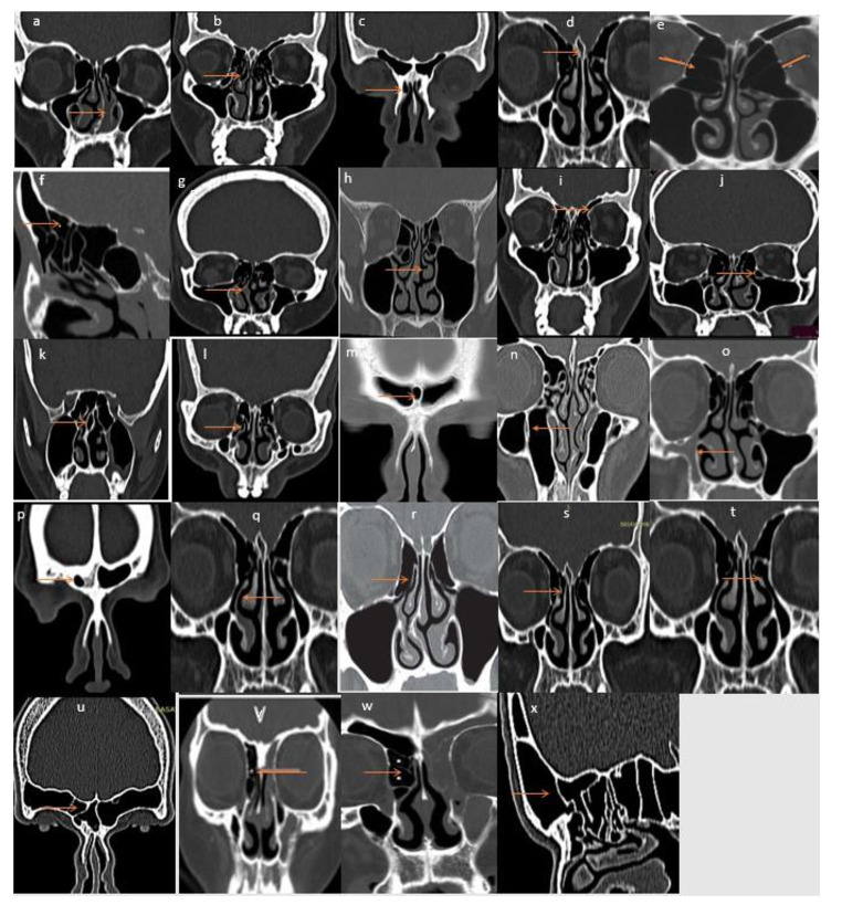
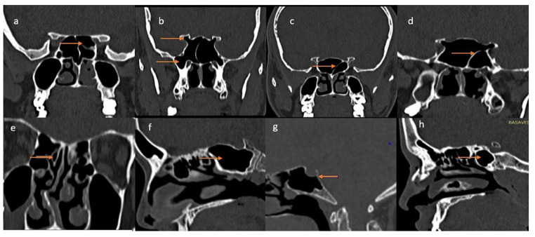
Results: All 70 CRS patients evaluated by CT PNS had one or more anatomical variations along with other findings like fungal sinusitis in 6 patients, dentigerous cyst and inverted papilloma in 1 case each. The anatomical variations observed in our study were septal deviation(62.8%), concha bullosa(52.8%), agger nasi(51.4%), pneumatized crista galli(47.1%), hyperpneumatized bulla(40%), suprabullar cells(37%), septal spur(34.3%), paradoxically curved middle turbinate(34.2%), supra orbital cells(32.8%), haller cells(31.4%), septal pneumatization(17.1%), pneumatized uncinate(13%), interfrontal septal pneumatization(13%), maxillary septations(10%), maxillary sinus hypoplasia(7.1%), frontal hypoplasia(5.7%), uncinate attached to lamina papyracea(40%), uncinate to middle turbinate(11%), uncinate to skull base(7.9%), free uncinate(41%), frontal cells type 1;2;3;4 (36%);(30%);(20%);(38.5%), onodi cells(27.1%), pneumatized anterior clinoid process(18.5%), lateral recess(15.7%), sphenoid septations attached to optic nerve(10%) and carotid(2.8%), pneumatized superior turbinate(1.4%), Sellar; Pre sellar; Post sellar sphenoid(42.8%);(5.7%);(51.4%). In our study only anatomical variations around the maxillary & Frontal sinus showed significant association with CRS.
Conclusion: Anatomical variations around the anterior group of sinuses have a significant association with CRS.
Keywords: Anatomical Variation; CT Paranasal Sinuses; Chronic Rhinosinusitis; EPOS 2020.
© Association of Otolaryngologists of India 2023. Springer Nature or its licensor (e.g. a society or other partner) holds exclusive rights to this article under a publishing agreement with the author(s) or other rightsholder(s); author self-archiving of the accepted manuscript version of this article is solely governed by the terms of such publishing agreement and applicable law.
Conflict of interest statement
Competing InterestsThe authors declare that they have no competing interest.Conflict of InterestThere is no conflict of interest.
Figures
Similar articles
-
Anatomical Variations of the Nose and Paranasal Sinuses: A Computed Tomographic Study.Indian J Otolaryngol Head Neck Surg. 2019 Nov;71(Suppl 3):2231-2240. doi: 10.1007/s12070-019-01716-9. Epub 2019 Jul 26. Indian J Otolaryngol Head Neck Surg. 2019. PMID: 31763326 Free PMC article.
-
Clinical and Radiological Significance of Anatomical Variations in Paranasal Sinuses: A Retrospective CT-Based Study.Cureus. 2025 Apr 18;17(4):e82506. doi: 10.7759/cureus.82506. eCollection 2025 Apr. Cureus. 2025. PMID: 40385872 Free PMC article.
-
The Anatomic Variations of the Nose and Paranasal Sinuses and Their Effect on Chronic Rhinosinusitis in Adult Patients.Indian J Otolaryngol Head Neck Surg. 2022 Oct;74(Suppl 2):960-966. doi: 10.1007/s12070-020-01975-x. Epub 2020 Aug 9. Indian J Otolaryngol Head Neck Surg. 2022. PMID: 36452856 Free PMC article.
-
Anatomical Variations of the Nasal Cavities and Paranasal Sinuses: A Systematic Review.Cureus. 2021 Jan 15;13(1):e12727. doi: 10.7759/cureus.12727. Cureus. 2021. PMID: 33614330 Free PMC article. Review.
-
Remarkable anatomic variations in paranasal sinus region and their clinical importance.Eur J Radiol. 2004 Jun;50(3):296-302. doi: 10.1016/j.ejrad.2003.08.012. Eur J Radiol. 2004. PMID: 15145491 Review.
References
-
- Carl Philpott. rhinosinusitis: definitions, classification and diagnosis. scott brown’s s otorhinolaryngology and head and neck surgery: basic sciences, endocrine surgery, rhinology 8th edi 2018 pages 1025–1034
-
- Tanvi rekhade AZ, nitnaware, Seema patel RT, Pawar Ashish Keche clinical profile of chronic rhinosinusitis: a study in central India. Indian J Res | Volume – 10 | Issue – 04 |April – 2021
-
- Wytske J, Fokkens VJ, Lund C, Hopkins PW, Hellings R, Kern S, Reitsma et al European position paper on Rhinosinusitis and nasal polyps 2020. Rhinol Suppl 29 vol 58: 1–464
-
- Wenrol Espinosa R, Genito, Rachel Zita Ramos Anatomic variations of the nasal cavity and paranasal sinus and their correlation with chronic rhinosinusitis using Harvard staging system. J Otolaryngol ENT Res. 2018;10(4):190–193.
-
- Rajneesh R (2017jul) Study of anatomical variations in osteomeatal complex in patients with chronic rhinosinusistis. Int J Otorhinolaryngol Head Neck Surg 3(3):528–533
LinkOut - more resources
Full Text Sources