

Morphometric Analysis of Temporal Bone Radiology for Cochlear Implant Candidacy
- PMID: 38440533
- PMCID: PMC10908920
- DOI: 10.1007/s12070-023-04257-4
Morphometric Analysis of Temporal Bone Radiology for Cochlear Implant Candidacy
Abstract
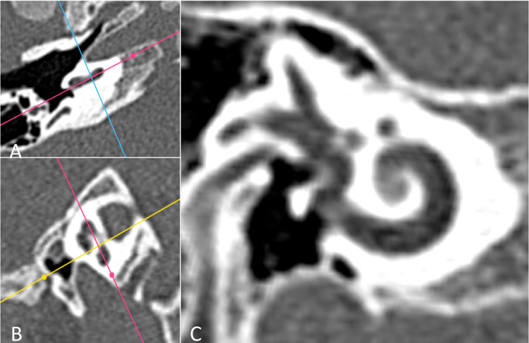
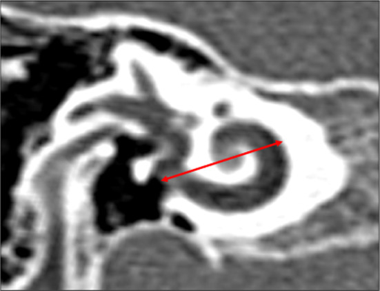
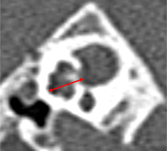
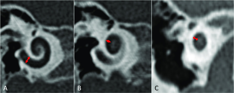
Cochlear Implantation (CI) is a well-accepted treatment for severe-to-profound sensorineural hearing loss, refractory to conventional hearing amplification. Pre-operative Computed Tomography (CT) and Magnetic Resonance Imaging (MRI) play pivotal roles in patient selection to rule out findings that preclude surgery or identify conditions that may impact the surgical procedure. A prospective study was carried out in a tertiary care center over three years, from January 2020 to January 2023. One hundred and ninety (380 ears) patients' High-Resolution Computed Tomography (HRCT) studies of the temporal bone and MRI scans of the auditory pathways were analyzed. A reporting format was followed which was devised by a team of senior implant surgeons and senior neuro-radiologists. Our study aims to provide a comprehensive radiologic protocol for CI candidacy including normative data for the essential morphometrics in the Indian setting.
Keywords: Cochlear implant; Morphometrics; Radiologic protocol.
© Association of Otolaryngologists of India 2023. Springer Nature or its licensor (e.g. a society or other partner) holds exclusive rights to this article under a publishing agreement with the author(s) or other rightsholder(s); author self-archiving of the accepted manuscript version of this article is solely governed by the terms of such publishing agreement and applicable law.
Conflict of interest statement
Conflict of interestThe authors report no conflict of interest.
Figures
Similar articles
-
Pediatric Single-Sided Deafness: A Review of Prevalence, Radiologic Findings, and Cochlear Implant Candidacy.Ann Otol Rhinol Laryngol. 2022 Mar;131(3):233-238. doi: 10.1177/00034894211019519. Epub 2021 May 26. Ann Otol Rhinol Laryngol. 2022. PMID: 34036833
-
Role of HRCT and MRI of the Temporal Bone in Predicting and Grading the Degree of Difficulty of Cochlear Implant Surgery.Indian J Otolaryngol Head Neck Surg. 2015 Jun;67(2):150-8. doi: 10.1007/s12070-015-0858-z. Epub 2015 May 5. Indian J Otolaryngol Head Neck Surg. 2015. PMID: 26075170 Free PMC article.
-
Imaging Modality of Choice for Pre-Operative Cochlear Imaging: HRCT vs. MRI Temporal Bone.J Clin Diagn Res. 2016 Oct;10(10):TC01-TC04. doi: 10.7860/JCDR/2016/18033.8592. Epub 2016 Oct 1. J Clin Diagn Res. 2016. PMID: 27891421 Free PMC article.
-
Cochlear implantation in children with anomalous cochleovestibular anatomy.Laryngoscope. 2005 Jan;115(1 Pt 2 Suppl 106):1-26. doi: 10.1097/00005537-200501001-00001. Laryngoscope. 2005. PMID: 15626926 Review.
-
Aspects of temporal bone anatomy and pathology in conjunction with cochlear implant surgery.Acta Radiol Suppl. 2003 Jul;430:2-15. Acta Radiol Suppl. 2003. PMID: 12834396 Review.
References
-
- Casselman JW, Veillon F. Temporal bone and auditory pathways. In: Hodler J, von Schulthess GK, Zollikofer CL, editors. Diseases of the brain, head & neck, Spine 2012–2015. Milano: Springer; 2012.
LinkOut - more resources
Full Text Sources