Literature Review, Case Presentation and Management of Non-ossifying Fibroma of Right Angle of Mandible: More Than just a Cortical Defect!
- PMID: 38440574
- PMCID: PMC10908682
- DOI: 10.1007/s12070-023-04110-8
Literature Review, Case Presentation and Management of Non-ossifying Fibroma of Right Angle of Mandible: More Than just a Cortical Defect!
Abstract
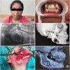
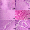
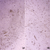
Non-ossifying fibroma (NOF) of jaw bones are rare. While NOF is the most common benign bone tumor of long bones with pathognomonic radiological features and bear a tendency for self-regression, gnathic NOF appears to be comparatively larger in size and behave more aggressively. A 16 years old female patient reported with painless swelling of the right side of the face of 4 months duration. Radiographic analysis showed a unilocular radiolucent lesion of right angle of the mandible with ill-defined margins, cortical perforation and thinning of inferior border. The lesion was provisionally diagnosed as odontogenic keratocyst/unicystic ameloblastoma and incisional biopsy was performed. The histopathological features and immunohistochemical characteristics favored a diagnosis of NOF. The lesion was excised and reconstructed. The excised specimen confirmed the diagnosis. There are no signs of recurrence at 18 months follow-up. NOF should be considered in the differential diagnosis of uni-/multilocular radiolucencies of jaws particularly the posterior mandible.
Keywords: ER; FGFR1; Gnathic; Mandible; Non-ossifying fibroma; RAS.
© Association of Otolaryngologists of India 2023. Springer Nature or its licensor (e.g. a society or other partner) holds exclusive rights to this article under a publishing agreement with the author(s) or other rightsholder(s); author self-archiving of the accepted manuscript version of this article is solely governed by the terms of such publishing agreement and applicable law.
Conflict of interest statement
Conflict of interestThe authors declare that they have no conflict of interest.
Figures


Similar articles
-
Bilateral Nonossifying Fibroma of the Mandible: A case report of a rare entity.Int J Mycobacteriol. 2023 Apr-Jun;12(2):196-206. doi: 10.4103/ijmy.ijmy_53_23. Int J Mycobacteriol. 2023. PMID: 37338484
-
Non-ossifying Fibroma of Mandible in a Four-Year-Old Girl: A Case Report.Cureus. 2023 Mar 21;15(3):e36470. doi: 10.7759/cureus.36470. eCollection 2023 Mar. Cureus. 2023. PMID: 37090356 Free PMC article.
-
The non-ossifying fibroma: a case report and review of the literature.Head Neck Pathol. 2013 Jun;7(2):203-10. doi: 10.1007/s12105-012-0399-7. Epub 2012 Sep 25. Head Neck Pathol. 2013. PMID: 23008139 Free PMC article. Review.
-
Amyloid Variant of Central Odontogenic Fibroma in the Mandible: A Case Report and Literature Review.Am J Case Rep. 2020 Aug 30;21:e925165. doi: 10.12659/AJCR.925165. Am J Case Rep. 2020. PMID: 32862189 Free PMC article. Review.
-
Maxillary unicystic ameloblastoma: a case report.BMC Res Notes. 2016 Oct 18;9(1):469. doi: 10.1186/s13104-016-2260-7. BMC Res Notes. 2016. PMID: 27756334 Free PMC article.
References
-
- WHO Classification of Tumours Editorial Board . Soft tissue and bone tumours. WHO classification of tumours series. 5. Lyon: International Agency for Research on Cancer; 2020. pp. 447–448.
-
- Sontag LW, Pyle DI. The appearance and nature of cyst-like areas in distal femoral metaphyses of children. Am J Roentgenol Radiat Ther. 1941;46:185–188.
LinkOut - more resources
Full Text Sources
Miscellaneous
