. 2024 Feb;76(1):1255-1259.
doi: 10.1007/s12070-023-04252-9. Epub 2023 Oct 15.

Gorlin-Goltz Syndrome: An Incidental Finding of a Rare Entity

Affiliations

Gorlin-Goltz Syndrome: An Incidental Finding of a Rare Entity

Sugandha Verma et al. Indian J Otolaryngol Head Neck Surg. 2024 Feb.

Abstract

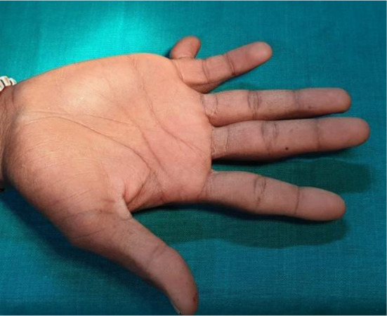
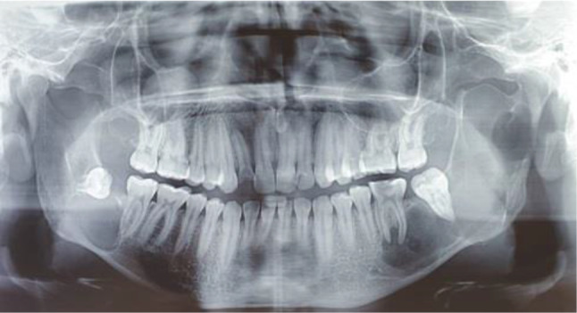
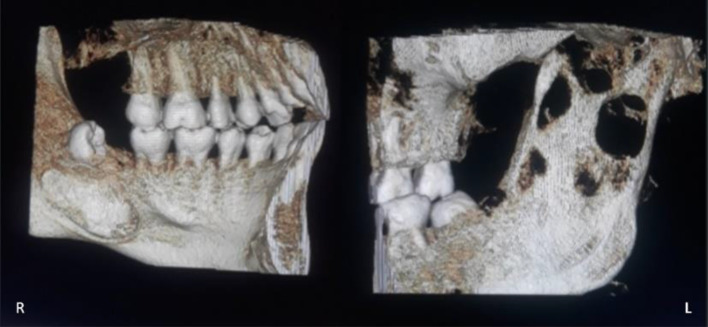
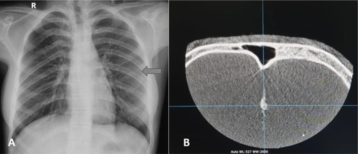
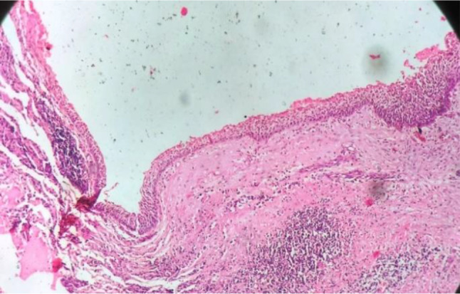
Gorlin-Goltz syndrome (GGS) is a rare hereditary disease characterized by multiple basal cell carcinomas, odontogenic keratocyst (OKCs) and musculoskeletal malformations. Pathogenesis of the syndrome is attributed to abnormalities in the long arm of chromosome 9 (q22.3-q31) and mutations in the human patched gene (PTCH1 gene). Here, we report a rare case of an incidental finding of GGS in an 18-year-old male patient presenting multiple OKCs, calcification of the falx cerebri, and bifid rib.

Keywords: Bifid rib; Gorlin–Goltz syndrome; Keratocystic odontogenic tumor; Odontogenic keratocyst.


Conflict of interest statement

Conflict of interestThe authors declare that they have no competing interests.

Figures

Fig. 1
Fig. 1
Intra-Oral image showing a carious tooth i.r.t 36
Fig. 2
Fig. 2
Polydactyly in the right hand
Fig. 3
Fig. 3
Orthopantomograph revealing osteolytic lesions on the right side of the maxilla and on the bilateral sides of the mandible. Root resorption is seen i.r.t 38 and 47. Mesio-angularly impacted tooth i.r.t 48
Fig. 4
Fig. 4
Image showing CBCT reconstruction of the lesions of the jaw
Fig. 5
Fig. 5
A Chest radiograph showing the presence of a bifid rib on the left side. B Computed tomography scan of the skull showing calcification of the falx cerebri
Fig. 6
Fig. 6
Photomicrograph showing a cystic lumen lined by wavy para-keratinized epithelium of uniform thickness supported by connective tissue capsule. The lumen is seen filled with keratin debris
Fig. 7
Fig. 7
Orthopantamograph of 6 months follow-up

Similar articles

References

    1. Pol CA, Ghige SK, Kalaskar RR, Gosavi SR. Gorlin–Goltz syndrome: a rare case report. Contemp Clin Dent. 2013;4:547–550. doi: 10.4103/0976-237X.123085. - DOI - PMC - PubMed
    1. Agrawal A, Murari A, Vutukuri S, Singh A. Gorlin–Goltz syndrome: case report of a rare hereditary disorder. Case Rep Dent. 2012;2012:4. doi: 10.1155/2012/47543. - DOI - PMC - PubMed
    1. Patil K, Mahima VG, Gupta B. Gorlin syndrome: a case report. J Indian Soc Pedod Prev Dent. 2005;23:198–203. doi: 10.4103/0970-4388.19010. - DOI - PubMed
    1. Kimonis VE, Goldstein AM, Pastakia B, Yang ML, Kase R, DiGiovanna JJ, et al. Clinical manifestations in 105 persons with nevoid basal cell carcinoma syndrome. Am J Med Genet. 1997;69:299–308. doi: 10.1002/(SICI)1096-8628(19970331)69:3<299::AID-AJMG16>3.0.CO;2-M. - DOI - PubMed
    1. Manfredi M, Vescovi P, Bonanini M, Porter S. Nevoid basal cell carcinoma syndrome: a review of the literature. Int J Oral Maxillofac Surg. 2004;33:117–124. doi: 10.1054/ijom.2003.0435. - DOI - PubMed

LinkOut - more resources