

A Detailed Assessment of Variations of Ethmoid Roof, Olfactory Fossa, and Anterior Ethmoidal Artery on CT Scan of Paranasal Sinuses of 200 Patients
- PMID: 38440628
- PMCID: PMC10909058
- DOI: 10.1007/s12070-023-04116-2
A Detailed Assessment of Variations of Ethmoid Roof, Olfactory Fossa, and Anterior Ethmoidal Artery on CT Scan of Paranasal Sinuses of 200 Patients
Abstract
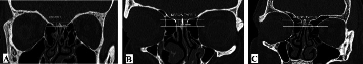
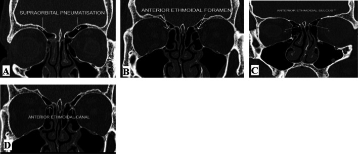
To study and analyse the variations in ethmoid roof anatomy and estimate the anatomical location and variations of AEA on CT scans. The study is conducted on 200 patients for detailed analysis of the olfactory fossa (OF) depth, supraorbital pneumatisation, and AEA location and distance from the skull base. In our study, Keros type II was predominant type seen followed by type I. Asymmetry was noted in 32/200 subjects (16%). The anterior ethmoidal artery (AEA) canal was seen in 341/400 sides (85.2%). We found Keros type II was the most common type in our study. We also found grade I anterior ethmoidal artery as the most common variant and the dangerous grade III anterior ethmoidal artery was least common type found in this study, and there was a significant association of Keros type II with increasing anterior ethmoidal artery grading.
Keywords: Anatomical variations; Anterior ethmoid artery; CT scan; Ethmoid roof; Olfactory fossa; Paranasal sinuses.
© Association of Otolaryngologists of India 2023. Springer Nature or its licensor (e.g. a society or other partner) holds exclusive rights to this article under a publishing agreement with the author(s) or other rightsholder(s); author self-archiving of the accepted manuscript version of this article is solely governed by the terms of such publishing agreement and applicable law.
Conflict of interest statement
Conflict of interestThe authors have not disclosed any competing interests.
Figures
References
-
- Jacob TG, Kaul JM. Morphology of the olfactory fossa—a new look. J Anatomic Soc India. 2014;63:30–35. doi: 10.1016/j.jasi.2014.04.006. - DOI
-
- Abdullah B, Lim EH, Husain S, Snidvongs K, Wang Y. Anatomical variations of anterior ethmoidal artery and their significance in endoscopic sinus surgery: a systematic review. Surg RadiolAnat. 2019;41:491–499. - PubMed
LinkOut - more resources
Full Text Sources
Research Materials