. 2024 Feb;76(1):224-236.
doi: 10.1007/s12070-023-04131-3. Epub 2023 Aug 8.

Subtotal Petrosectomy: Pictorial Review of Clinical Indications and Surgical Approach

Affiliations

Subtotal Petrosectomy: Pictorial Review of Clinical Indications and Surgical Approach

Riccardo Nocini et al. Indian J Otolaryngol Head Neck Surg. 2024 Feb.

Abstract

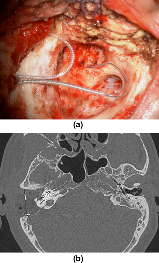
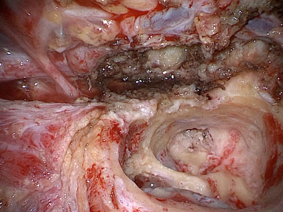
Subtotal petrosectomy (STP) is characterized by obliteration of the middle ear and occlusion of the external auditory canal. The advent of the endoscope has allowed a reduction in morbidity for some conditions such as cholesteatoma and other middle ear disorders, but STP still plays an important role. A retrospective review of medical records and videos of patients who had undergone STP was performed. Perioperative data and images were collected from various clinical cases who had undergone subtotal petrosectomy at our tertiary referral university hospital in Verona. We confronted our experience with a review of the literature to present the main indications for this type of procedure. STP allows a variety of diseases to be managed effectively as it offers the possibility of a definitive healing with radical clearance of temporal bone. Moreover, it can be safely combined with other procedures with a very low complication rate. Although the endoscope represents a revolution in ear surgery, STP, when indicated, is nowadays a surgical option that should be included in the otosurgeon's portfolio.

Keywords: Cholesteatoma; Cochlear implant; Skull base tumors; Subtotal petrosectomy; Temporal bone fractures.


Conflict of interest statement

Conflict of interestAll the authors declare that they have no conflict of interest.

Figures

Fig. 1
Fig. 1
a, b EAC eversion and “blind sac closure”; c postoperative view with completely drilled air cells; d obliteration of the cavity with abdominal fat and closure of the Eustachian tube (ET) with a fragment of the temporal muscle
Fig. 2
Fig. 2
a, b Preoperative CT scan: axial view and coronal view with hypodense material in the left tympanic cavity and in the left mastoid cells; erosion of the left tegmen tympani is clearly seen (*); c intraoperative view: mastoid cholesteatoma; d intraoperative view: complete drilling of the air cells including the retrofacial; no evidence of residual cholesteatoma; a large tegmental defect is visible (*)
Fig. 3
Fig. 3
a CT scan, axial, right ear: a giant cholesteatoma occupies the middle ear and erodes the surrounding structures. A CSL fistula can be seen; b preoperative CT scan: axial view with left tegmen tympani dehiscence (arrow); c intraoperative view: large left tegmen tympani and mastoideum dehiscence; d post-operative MRI: left petrosectomy with abdominal fat in neo-cavity (*)
Fig. 4
Fig. 4
a Preoperative CT scan: right temporal bone fracture witch cross toward the vestibule; b, c intraoperative view: facial decompression with epineurium section from the geniculate ganglion to the stylomastoid foramen. In this particular case, given the fact the trauma caused the deaf ear of this side, we chose to perform a partial labyrinthectomy (drilling of lateral semicircular canal) to decompress full the second genu of the facial nerve
Fig. 5
Fig. 5
a Preoperative CT scan: axial view with ipodense material in the left middle ear and mastoid cells; b maxillary artery angiography: contrast blush(*) at the level of the inferior part of the left mastoid bone, compatible with tympanic glomus, is documented; c, d intraoperative view: the highly vascularization of the paraganglioma and its subsequent bleeding during dissection worsen the visualization of the anatomical landmarks
Fig. 6
Fig. 6
a Intraoperative view after right CI placement. Removal of the posterior wall of the EAC improves visualization of the round window in difficult anatomical conditions; b postoperative CT scan: the array is correctly placed in the right cochlea; complete air cells drilling with ipodense material in the neo-cavity (abdominal fat)
Fig. 7
Fig. 7
a Preoperative CT scan: the left IAC has a tapered scape and incomplete partition type 3. Note the fundus of the internal auditory canal, which communicates with the basal turn of the cochlea and is not separated by bone (arrow). In this extreme case, we performed an STP because the risk of gusher was too high and to better control the insertion of the array; b, c intraoperative view: the array is inserted; in this extreme case the cochlea was partially drilled (infrapromontorial approach) to visualize the basal turn of the cochlea and to ensure that it is not inserted into the internal auditory canal by dehiscence of the medial wall of the cochlea; d postoperative CT scan: the array is correctly placed in the left cochlea; complete air cells drilling with ipodense material in the neo-cavity (*)
Fig. 8
Fig. 8
Intraoperative view: petrosectomy for a malignancy of the EAC; the facial nerve is seen from the geniculate ganglion to its entrance in the parotid gland
Fig. 9
Fig. 9
a, b Even after STP, recurrence may occur. In this MRI image, coronal planes, we see the left middle ear filled with fat and a hypointense round lesion. This was confirmed as a cholesteatoma in the DWI sequences

References

    1. Marchioni D, Gazzini L, De Rossi S, Di Maro F, Sacchetto L, Carner M, Bianconi L. The management of tympanic membrane perforation with endoscopic type I tympanoplasty. Otol Neurotol. 2020;41(2):214–221. doi: 10.1097/MAO.0000000000002465. - DOI - PubMed
    1. Marchioni D, Gazzini L, Bonali M, Bisi N, Presutti L, Rubini A. Role of endoscopy in lateral skull base approaches to the petrous apex. Eur Arch Otorhinolaryngol. 2020;277(3):727–733. doi: 10.1007/s00405-019-05750-9. - DOI - PubMed
    1. Marchioni D, Rubini A, Nogueira JF, Isaacson B, Presutti L. Transcanal endoscopic approach to lesions of the suprageniculate ganglion fossa. Auris Nasus Larynx. 2018;45(1):57–65. doi: 10.1016/j.anl.2017.03.006. - DOI - PubMed
    1. Coker NJ, Jenkins HA, Fisch U. Obliteration of the middle ear and mastoid cleft in subtotal petrosectomy: indications, technique, and results. Ann Otol Rhinol Laryngol. 1986;95(1 Pt 1):5–11. doi: 10.1177/000348948609500102. - DOI - PubMed
    1. Fisch U. Microsurgery of the skull base. Stuttgart: Georg Thieme Verlag; 1988.

LinkOut - more resources