

Relation Between Reactive Oxygen Species Production and Transient Receptor Potential Vanilloid1 Expression in Human Skin During Aging
- PMID: 38440794
- PMCID: PMC10956443
- DOI: 10.1369/00221554241236537
Relation Between Reactive Oxygen Species Production and Transient Receptor Potential Vanilloid1 Expression in Human Skin During Aging
Abstract
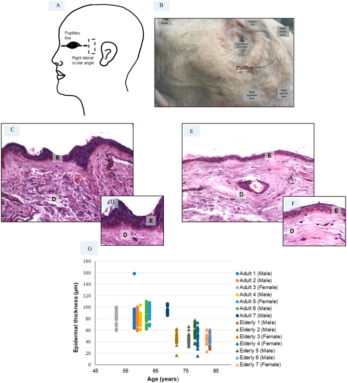
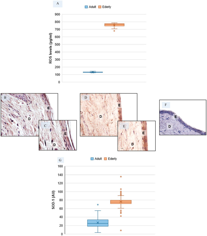
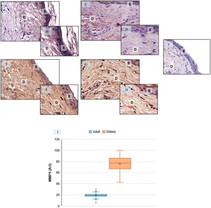
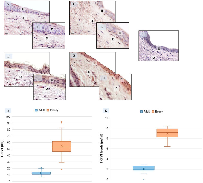
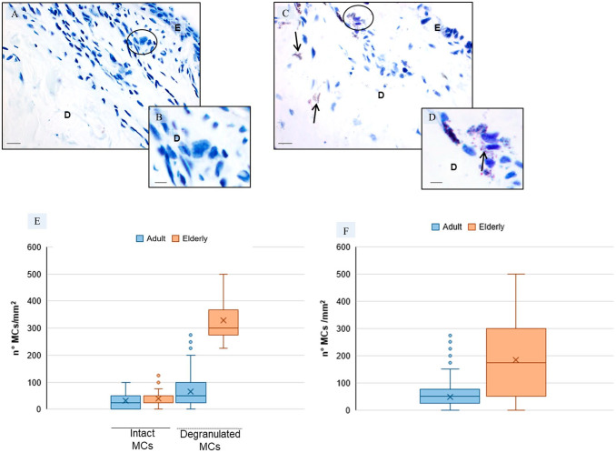
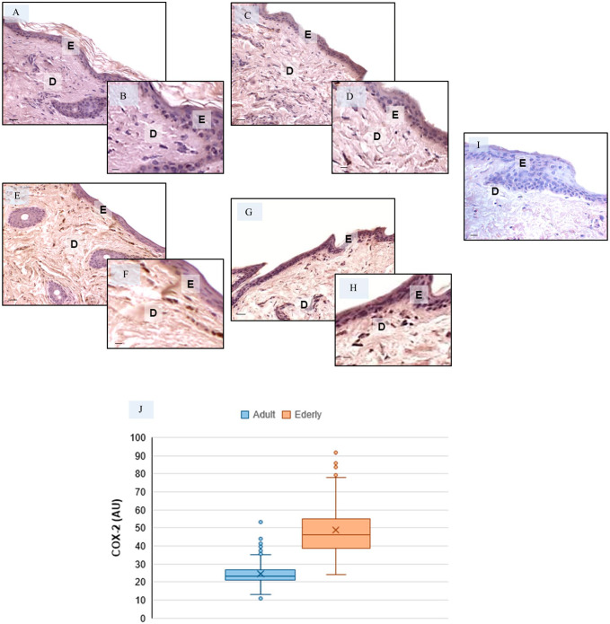
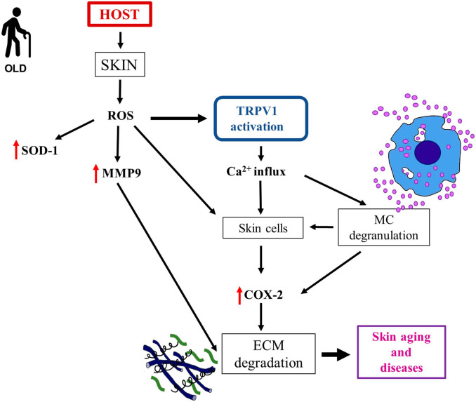
Skin sensitivity and impaired epidermal barrier function are associated with aging and are at least partly due to increased production of reactive oxygen species (ROS). Transient receptor potential vanilloid1 (TRPV1) is expressed in keratinocytes, fibroblasts, mast cells, and endothelial cells in skin. We investigated in skin biopsies of adult and elderly donors whether TRPV1 expression is involved in the skin aging process. We found that aging skin showed a strongly reduced epidermal thickness, strongly increased oxidative stress, protease expression, and mast cell degranulation and strongly increased TRPV1 expression both in epidermis and dermis. Based on our findings, the aging-related changes observed in the epidermis of the skin level are associated with increased ROS production, and hypothesized alterations in TRPV1 expression are mechanistically linked to this process.
Keywords: aging; cyclooxygenase-2; metalloproteinases; oxidative stress; skin; transient receptor potential vanilloid1 (TRPV1).
Conflict of interest statement
Competing InterestsThe author(s) declared no potential conflicts of interest with respect to the research, authorship, and/or publication of this article.
Figures
Similar articles
-
Paclitaxel-Induced Epidermal Alterations: An In Vitro Preclinical Assessment in Primary Keratinocytes and in a 3D Epidermis Model.Int J Mol Sci. 2022 Jan 20;23(3):1142. doi: 10.3390/ijms23031142. Int J Mol Sci. 2022. PMID: 35163066 Free PMC article.
-
Triolein reduces MMP-1 upregulation in dermal fibroblasts generated by ROS production in UVB-irradiated keratinocytes.J Dermatol Sci. 2017 Feb;85(2):124-130. doi: 10.1016/j.jdermsci.2016.11.010. Epub 2016 Nov 27. J Dermatol Sci. 2017. PMID: 27932255
-
The role of TRPV1 channel in aged human skin.J Dermatol Sci. 2012 Feb;65(2):81-5. doi: 10.1016/j.jdermsci.2011.11.003. Epub 2011 Nov 18. J Dermatol Sci. 2012. PMID: 22154816 Review.
-
Transient receptor potential vanilloid 1 plays a major role in low temperature-mediated skin barrier dysfunction.J Allergy Clin Immunol. 2022 Aug;150(2):362-372.e7. doi: 10.1016/j.jaci.2022.01.027. Epub 2022 Feb 19. J Allergy Clin Immunol. 2022. PMID: 35189126
-
[Human skin reconstructed in vitro as a model to study the keratinocyte, the fibroblast and their interactions: photodamage and repair processes].J Soc Biol. 2005;199(4):313-20. doi: 10.1051/jbio:2005032. J Soc Biol. 2005. PMID: 16738525 Review. French.
Cited by
-
Research Progress in Skin Aging and Immunity.Int J Mol Sci. 2024 Apr 7;25(7):4101. doi: 10.3390/ijms25074101. Int J Mol Sci. 2024. PMID: 38612909 Free PMC article. Review.
-
Skin Aging and the Upcoming Role of Ferroptosis in Geroscience.Int J Mol Sci. 2024 Jul 28;25(15):8238. doi: 10.3390/ijms25158238. Int J Mol Sci. 2024. PMID: 39125810 Free PMC article. Review.
-
Melatonin Attenuates Ferritinophagy/Ferroptosis by Acting on Autophagy in the Liver of an Autistic Mouse Model BTBR T+Itpr3tf/J.Int J Mol Sci. 2024 Nov 23;25(23):12598. doi: 10.3390/ijms252312598. Int J Mol Sci. 2024. PMID: 39684310 Free PMC article.
-
Organoids as Tools for Investigating Skin Aging: Mechanisms, Applications, and Insights.Biomolecules. 2024 Nov 12;14(11):1436. doi: 10.3390/biom14111436. Biomolecules. 2024. PMID: 39595612 Free PMC article. Review.
References
Publication types
MeSH terms
Substances
LinkOut - more resources
Full Text Sources
Research Materials
