. 2024 May 1;326(5):L574-L588.
doi: 10.1152/ajplung.00018.2024. Epub 2024 Mar 5.

P. aeruginosa tRNA-fMet halves secreted in outer membrane vesicles suppress lung inflammation in cystic fibrosis

Affiliations

P. aeruginosa tRNA-fMet halves secreted in outer membrane vesicles suppress lung inflammation in cystic fibrosis

Zhongyou Li et al. Am J Physiol Lung Cell Mol Physiol. .

Abstract

Although tobramycin increases lung function in people with cystic fibrosis (pwCF), the density of Pseudomonas aeruginosa (P. aeruginosa) in the lungs is only modestly reduced by tobramycin; hence, the mechanism whereby tobramycin improves lung function is not completely understood. Here, we demonstrate that tobramycin increases 5' tRNA-fMet halves in outer membrane vesicles (OMVs) secreted by laboratory and CF clinical isolates of P. aeruginosa. The 5' tRNA-fMet halves are transferred from OMVs into primary CF human bronchial epithelial cells (CF-HBEC), decreasing OMV-induced IL-8 and IP-10 secretion. In mouse lungs, increased expression of the 5' tRNA-fMet halves in OMVs attenuated KC (murine homolog of IL-8) secretion and neutrophil recruitment. Furthermore, there was less IL-8 and neutrophils in bronchoalveolar lavage fluid isolated from pwCF during the period of exposure to tobramycin versus the period off tobramycin. In conclusion, we have shown in mice and in vitro studies on CF-HBEC that tobramycin reduces inflammation by increasing 5' tRNA-fMet halves in OMVs that are delivered to CF-HBEC and reduce IL-8 and neutrophilic airway inflammation. This effect is predicted to improve lung function in pwCF receiving tobramycin for P. aeruginosa infection.NEW & NOTEWORTHY The experiments in this report identify a novel mechanism, whereby tobramycin reduces inflammation in two models of CF. Tobramycin increased the secretion of tRNA-fMet halves in OMVs secreted by P. aeruginosa, which reduced the OMV-LPS-induced inflammatory response in primary cultures of CF-HBEC and in mouse lung, an effect predicted to reduce lung damage in pwCF.

Keywords: P. aeruginosa; cystic fibrosis; host-pathogen; outer membrane vesicles; tRNA.


Conflict of interest statement

No conflicts of interest, financial or otherwise, are declared by the authors.

Figures

None
Graphical abstract
Figure 1.
Figure 1.
A and B: the LIVE/DEAD BacLight Viability Kit revealed that tobramycin (1 µg/mL) reduced the viability of P. aeruginosa. A t test was used to determine significance (**P = 0.0065 in A and **P = 0.0061 in B). The means are presented as red dots. Each black dot represents an experiment with a unique culture of PA14 exposed to vehicle or tobramycin on different days. C: growth curves of PA14 in the presence of vehicle or tobramycin (1 μg/mL; PA14 + Tobi) show significantly reduced growth of PA14 as determined by measuring OD600. The curves first significantly diverge at 1.15 h (P = 0.0141, t test) and remain significantly different thereafter, for example, P = 0.0017 at 2.00 h. Lines represent the averages from three biological replicates conducted of different days, and error bars (too small to see) indicate SE.
Figure 2.
Figure 2.
Nanosight particle tracking analysis (NTA) of OMVs isolated using the OptiPrep-Density Gradient. A: NTA of OMVs detected in each OptiPrep-fraction from six separate experiments conducted on different days. Vehicle-exposed (V-OMV) P. aeruginosa, and tobramycin-exposed P. aeruginosa (Tobi-OMV) are shown. The red dots indicate the means. The number of Tobi-OMVs was significantly above V-OMVs in fractions 2 and 3; thus, OMVs were pooled from fractions 2–3 from V-OMV, and fractions 2–3 were pooled from Tobi-OMV. n = 6 experiments conducted on separated days with different grow ups of P. aeruginosa. B: total counts of OMVs in fractions 2 and 3 combined from the same experiments depicted in A (n = 6). C: number of V-OMVs and Tobi-OMVs in 12 independent experiments conducted on different days where fractions 2 and 3 were combined for reported experiments. Each bacterial preparation was separated into two aliquotes, one was treated with vehicle and the other tobramycin, and then OMVs were isolated as described in METHODS. The lines connect experiments on the same day. *P =. 0306 in B, **P =. 0040 in C. OMV, outer membrane vesicle.
Figure 3.
Figure 3.
Representative transmission electron microscopy images of negatively stained OMV preparations and process control. The left-hand column shows images acquired at an original magnification of ×10,000 and the right-hand column shows the boxed areas from the left-hand column images acquired at an original magnification of ×30,000. Scale bars represent 500 nm and 100 nm for the low and high magnifications, respectively. Images of V-OMVs: low magnification (A) and high magnification (B). Images of Tobi-OMVs: low magnification (C) and high magnification (D). E and F: images showing the lack of identifiable vesicles in process control (media not exposed to PA14 and run through the OMV isolation procedure). Low magnification (E) and (F) high magnification. Experiments repeated twice on separate days. The electron microscopist was blinded to the treatment. OMV, outer membrane vesicle.
Figure 4.
Figure 4.
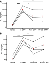
CF-HBECs from three donors were exposed to either vehicle, the same number of V-OMVs, or 40% more Tobi-OMVs, to represent the increased secretion of OMVs induced by tobramycin in the in vitro experiments. The means are presented as red dots. The concentration of OMVs used was in the range of bacterial vesicles measured in biological fluids, including BALF, in vivo (reviewed in Refs. 28, 29). After a 6-h exposure, the basolateral medium was collected, and cytokines were interrogated by 41-plex ELISA. Cytokines other than IL-8 and IP-10 were not significantly different between V-OMV and Tobi-OMV. Lines connect experiments conducted with CF-HBECs from the same donor on the same day. A: V-OMVs increased IL-8 secretion compared to vehicle (***P = 0.0007). Tobi-OMVs (1.5 × 10¹⁰ for a 12-mm filter) reduced IL-8 secretion compared to V-OMVs, (*P = 0.0341) as did 1.4 times as many Tobi-OMVs (2.1 × 10¹⁰ for a 12-mm filter) (*P =. 0334) B: V-OMVs significantly increased IP-10 secretion compared to vehicle (***P = 0.0004). Tobi-OMVs significantly reduced IP-10 secretion compared to V-OMVs (*P = 0.0218). Lines connect experiments conducted with CF-HBECs from the same donor. Linear mixed-effects models with CF-HBEC donor as a random effect were used to calculate P values. BALF, bronchoalveolar lavage fluid; CF-HBEC, CF human bronchial epithelial cell; ELISA, enzyme-linked immunosorbent assay; OMV, outer membrane vesicle.
Figure 5.
Figure 5.
Tobramycin increases the abundance of tRNA-fMet halves in OMVs. The input of small RNAs into the sequencing process was similar in each group (see METHODS). A: M vs. A plot (MA plot; M is the difference between the log intensity values and A is the average of the log intensity values), comparing the small RNA expression profile in V-OMVs and Tobi-OMVs (n = 3 for each group). Each dot represents a unique sequence read. The most abundant and most induced sRNAs in OMVs by tobramycin are highlighted in red and listed in Table 1. Length distribution of tRNA-fMet halves secreted in Tobi-OMVs mapped to gene locus PA14_62790 (B) and PA14_52320 (C). D: secondary cloverleaf structure of tRNA-fMet and cleavage site in the anticodon loop to generate tRNA-fMet halves. The red line indicates the only different pair of nucleotides between the two 5′ tRNA-fMet halves, and the red dot represents the only nucleotide difference between the two 5′ tRNA-fMet halves. E: qPCR for 5′ tRNA-fMet halves in V-OMVs and Tobi-OMVs purified from PA14 and four CF clinical isolates (n = 5 strains), including two mucoid and two nonmucoid CF clinical isolates. Tobi-OMV increased tRNA-fMet (*P = 0.0128, mixed effect linear model with strain as a random effect). The qPCR primers and probe were designed to detect both 5′ tRNA-fMet halves. The red dots indicate the mean. CF, cystic fibrosis; OMV, outer membrane vesicle.
Figure 6.
Figure 6.
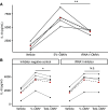
tRNA-fMet halves reduce IL-8 secretion in vitro. A: empty vector OMVs (EV-OMV) increased IL-8 secretion by CF-HBECs (n = 4 donors). OMVs overexpressing tRNA-fMet1 half (tRNA1-OMVs) reduced IL-8 secretion compared to EV-OMV (**P = 0.0055). B: the 1.4X Tobi-OMV effect of reducing IL-8 secretion was abolished (P = 0.4073) by transfection of an antisense RNA oligo inhibitor (tRNA1 inhibitor) in CF-HBEC that anneals to both tRNA-fMet halves but not by transfection of inhibitor negative control (n = 4 donors, *P = 0.0374). Lines in panels A and B connect data points using the cells from the same donor on the same day. The means are presented as red dots. A linear mixed-effects model with CF-HBEC donor as a random effect was used to calculate P values for data in A and B. CF-HBEC, CF human bronchial epithelial cell; OMV, outer membrane vesicle.
Figure 7.
Figure 7.
tRNA-fMet halves alter protein expression in CF-HBEC. A: volcano plot of proteomic analysis of polarized CF-HBECs (n = 3 donors) exposed to tRNA-fMet1 half (tRNA1-OMVs) compared to cells treated with EV-OMVs. The top 20% differentially expressed proteins are colored in blue (FDR P < 0.05 and >2-fold increase in abundance). Red dots with numbers represent downregulated proteins corresponding to proteins numbered in B. B: ingenuity pathway analysis (IPA) identified a downregulated proinflammatory network in five consensus pathways (Table 2), predicting decreased IL-8 expression. The green circles identify proteins whose abundance was reduced based on proteomic analysis. Blue shading indicates predicted inhibition. CF-HBEC, CF human bronchial epithelial cell; FDR, false discovery rate; OMV, outer membrane vesicle.
Figure 8.
Figure 8.
tRNA-fMet halves reduce IL-8 secretion and neutrophil recruitment in vivo. BALF from male and female mice exposed to EV-OMVs or tRNA1-fMet half OMVs was collected to measure KC concentration (A) and neutrophils (B). A: tRNA1-fMet half OMVs reduced KC (*P =. 01721, Wilcoxon rank-sum test) and (B) neutrophils (*P =. 02067, Wilcoxon rank-sum test). Nine to 10 mice were used per group. IL-8 in BALF was reduced in four pwCF during the 4-wk administration of inhaled tobramycin “On Tobi” (*P =. 0155) compared with off Tobi (C), and neutrophils in BALF from the same pwCF were reduced (*P =. 0035), while they were on Tobi compared with off Tobi (D). Lines connect data points from the same pwCF. Linear mixed-effect models were used to account for donor-to-donor variability and the number of days between collection dates for each sample pair (Supplemental Table S1). Red dots indicate the mean. Results were independent of the order of sample collection. The Dartmouth College IRB will not approve consecutive, prospective bronchoscopies in pwCF for research purposes; thus, samples were not collected in consecutive months. The samples collected and reported herein represent clinically justified samples of BALF to be used for research approved by the Dartmouth IRB. BALF, bronchoalveolar lavage fluid; OMV, outer membrane vesicle; pwCF, people with cystic fibrosis.

Update of

Similar articles

Cited by

References

    1. Stanton BA. Effects of Pseudomonas aeruginosa on CFTR chloride secretion and the host immune response. Am J Physiol Cell Physiol 312: C357–C366, 2017. doi: 10.1152/ajpcell.00373.2016. - DOI - PMC - PubMed
    1. Stoltz DA, Meyerholz DK, Welsh MJ. Origins of cystic fibrosis lung disease. N Engl J Med 372: 351–362, 2015. doi: 10.1056/NEJMra1300109. - DOI - PMC - PubMed
    1. Hauser AR, Jain M, Bar-Meir M, McColley SA. Clinical significance of microbial infection and adaptation in cystic fibrosis. Clin Microbiol Rev 24: 29–70, 2011. doi: 10.1128/CMR.00036-10. - DOI - PMC - PubMed
    1. Lin CK, Kazmierczak BI. Inflammation: a double-edged sword in the response to Pseudomonas aeruginosa infection. J Innate Immun 9: 250–261, 2017. doi: 10.1159/000455857. - DOI - PMC - PubMed
    1. Roesch EA, Nichols DP, Chmiel JF. Inflammation in cystic fibrosis: an update. Pediatr Pulmonol 53: S30–S50, 2018. doi: 10.1002/ppul.24129. - DOI - PubMed

Publication types

MeSH terms