

The cognitive relevance of non-lesional damage to cortical networks in people with multiple sclerosis
- PMID: 38441612
- PMCID: PMC11136718
- DOI: 10.1007/s00415-024-12240-4
The cognitive relevance of non-lesional damage to cortical networks in people with multiple sclerosis
Abstract
Background: Cognitive impairment, a common and debilitating symptom in people with multiple sclerosis (MS), is especially related to cortical damage. However, the impact of regional cortical damage remains poorly understood. Our aim was to evaluate structural (network) integrity in lesional and non-lesional cortex in people with MS, and its relationship with cognitive dysfunction.
Methods: In this cross-sectional study, 176 people with MS and 48 healthy controls underwent MRI, including double inversion recovery and diffusion-weighted scans, and neuropsychological assessment. Cortical integrity was assessed based on fractional anisotropy (FA) and mean diffusivity (MD) within 212 regions split into lesional or non-lesional cortex, and grouped into seven cortical networks. Integrity was compared between people with MS and controls, and across cognitive groups: cognitively-impaired (CI; ≥ two domains at Z ≤ - 2 below controls), mildly CI (≥ two at - 2 < Z ≤ - 1.5), or cognitively-preserved (CP).
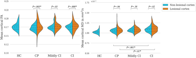
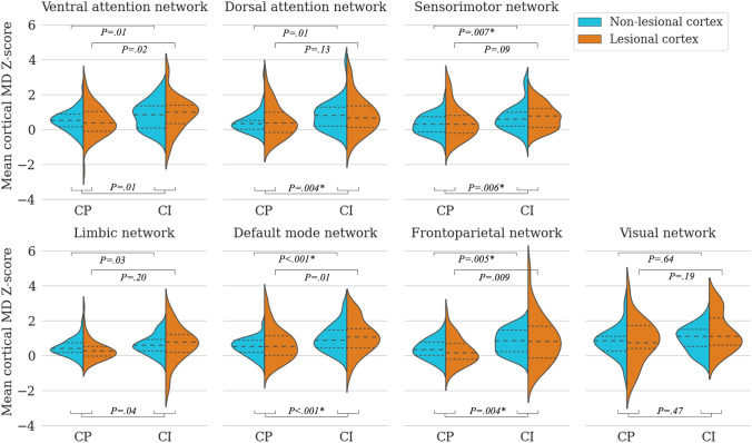
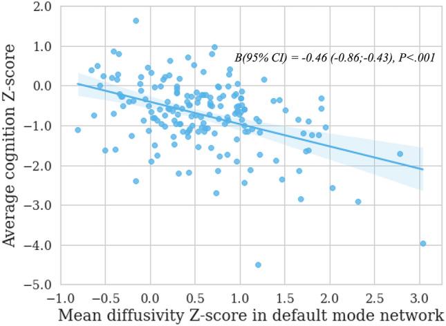
Results: Cortical lesions were observed in 87.5% of people with MS, mainly in ventral attention network, followed by limbic and default mode networks. Compared to controls, in non-lesional cortex, MD was increased in people with MS, but mean FA did not differ. Within the same individual, MD and FA were increased in lesional compared to non-lesional cortex. CI-MS exhibited higher MD than CP-MS in non-lesional cortex of default mode, frontoparietal and sensorimotor networks, of which the default mode network could best explain cognitive performance.
Conclusion: Diffusion differences in lesional cortex were more severe than in non-lesional cortex. However, while most people with MS had cortical lesions, diffusion differences in CI-MS were more prominent in non-lesional cortex than lesional cortex, especially within default mode, frontoparietal and sensorimotor networks.
Keywords: Cognition; Cortical lesions; Diffusion; Multiple sclerosis; Networks.
© 2024. The Author(s).
Conflict of interest statement
EAK, TAAB, and AB report no conflicts of interest; SN is supported by research grants from Atara Biotherapeutics, Merck and Biogen; MvD is supported by a research grant from BMS. PMB received research support from the Dutch MS Research Foundation; FB is a steering committee or iDMC member for Biogen, Merck, Roche, EISAI, Prothena, is a consultant to Roche, Biogen, Merck, IXICO, Jansen, Combinostics, has research agreements with Novartis, Merck, Biogen, GE Healthcare, and Roche, and is co-founder & shareholder of Queen Square Analytics LTD; BMJU reports research support and/or consultancy fees from Biogen Idec, Genzyme, Merck Serono, Novartis, Roche, Teva, and Immunic Therapeutics; ECK has received consulting fees from EMD Serono, Genentech, INmune Bio, Myrobalan Therapeutics, OM1and TG Therapeutics, and received research funds from Abbvie, Biogen, and Genentech; IK received research grants from LabEx TRAIL and ARSEP and speakers’ honoraria from Celgene; MMS serves on the editorial board of Neurology and Frontiers in Neurology and Multiple Sclerosis Journal, receives research support from the Dutch MS Research Foundation, Eurostars-EUREKA, ARSEP, Amsterdam Neuroscience, MAGNIMS and ZonMW and has served as a consultant for or received research support from EIP Pharma, Atara Biotherapeutics, Biogen, Celgene/Bristol Meyers Squibb, Genzyme, MedDay and Merck.
Figures

Similar articles
-
Cortical lesions impact cognitive decline in multiple sclerosis via volume loss of nonlesional cortex.Ann Clin Transl Neurol. 2025 Jan;12(1):121-136. doi: 10.1002/acn3.52261. Epub 2024 Dec 27. Ann Clin Transl Neurol. 2025. PMID: 39729590 Free PMC article.
-
Energy Associated With Dynamic Network Changes in Patients With Multiple Sclerosis and Cognitive Impairment.Neurology. 2024 Nov 12;103(9):e209952. doi: 10.1212/WNL.0000000000209952. Epub 2024 Oct 11. Neurology. 2024. PMID: 39393029 Free PMC article.
-
DT MRI microstructural cortical lesion damage does not explain cognitive impairment in MS.Mult Scler. 2017 Dec;23(14):1918-1928. doi: 10.1177/1352458516689147. Epub 2017 Jan 18. Mult Scler. 2017. PMID: 28098510
-
Cortical lesions and cognitive impairment in multiple sclerosis.Neurol Sci. 2010 Nov;31(Suppl 2):S235-7. doi: 10.1007/s10072-010-0368-4. Neurol Sci. 2010. PMID: 20635113 Review.
-
Cognitive impairment in multiple sclerosis: from phenomenology to neurobiological mechanisms.J Neural Transm (Vienna). 2024 Aug;131(8):871-899. doi: 10.1007/s00702-024-02786-y. Epub 2024 May 18. J Neural Transm (Vienna). 2024. PMID: 38761183 Review.
Cited by
-
In vivo evidence for cell body loss in cortical lesions in people with multiple sclerosis.Ann Clin Transl Neurol. 2025 Jan;12(1):4-16. doi: 10.1002/acn3.52237. Epub 2024 Dec 13. Ann Clin Transl Neurol. 2025. PMID: 39673156 Free PMC article.
-
Evolution of Cortical Lesions and Function-Specific Cognitive Decline in People With Multiple Sclerosis.Neurology. 2025 Jun;104(11):e213650. doi: 10.1212/WNL.0000000000213650. Epub 2025 May 12. Neurology. 2025. PMID: 40354586 Free PMC article.
References
-
- Sumowski JF, Benedict R, Enzinger C, Filippi M, Geurts JJ, Hamalainen P, Hulst H, Inglese M, Leavitt VM, Rocca MA, Rosti-Otajarvi EM, Rao S. Cognition in multiple sclerosis: state of the field and priorities for the future. Neurology. 2018;90:278–288. doi: 10.1212/WNL.0000000000004977. - DOI - PMC - PubMed
-
- Haider L, Prados F, Chung K, Goodkin O, Kanber B, Sudre C, Yiannakas M, Samson RS, Mangesius S, Thompson AJ, Gandini Wheeler-Kingshott CAM, Ciccarelli O, Chard DT, Barkhof F. Cortical involvement determines impairment 30 years after a clinically isolated syndrome. Brain. 2021;144:1384–1395. doi: 10.1093/brain/awab033. - DOI - PMC - PubMed
MeSH terms
Grants and funding
LinkOut - more resources
Full Text Sources
Medical
Miscellaneous
