. 2024 Apr 2;12(4):e0322023.
doi: 10.1128/spectrum.03220-23. Epub 2024 Mar 5.

Heme oxygenase-1 is an equid alphaherpesvirus 8 replication restriction host protein and suppresses viral replication via the PKCβ/ERK1/ERK2 and NO/cGMP/PKG pathway

Affiliations

Heme oxygenase-1 is an equid alphaherpesvirus 8 replication restriction host protein and suppresses viral replication via the PKCβ/ERK1/ERK2 and NO/cGMP/PKG pathway

Tongtong Wang et al. Microbiol Spectr. .

Abstract

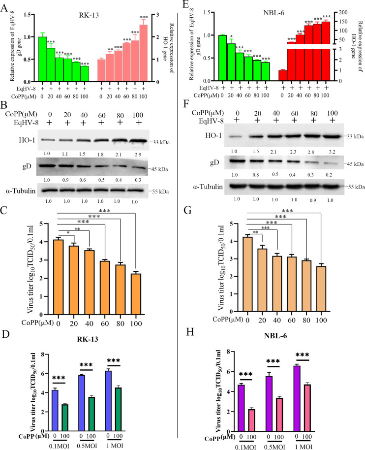
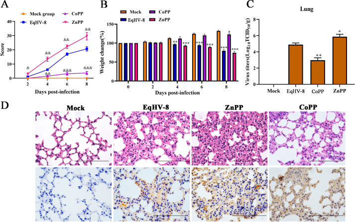
Equid alphaherpesvirus 8 (EqHV-8) is one of the most economically important viruses that is known to cause severe respiratory disease, abortion, and neurological syndromes in equines. However, no effective vaccines or therapeutic agents are available to control EqHV-8 infection. Heme oxygenase-1 (HO-1) is an antioxidant defense enzyme that displays significant cytoprotective effects against different viral infections. However, the literature on the function of HO-1 during EqHV-8 infection is little. We explored the effects of HO-1 on EqHV-8 infection and revealed its potential mechanisms. Our results demonstrated that HO-1 induced by cobalt-protoporphyrin (CoPP) or HO-1 overexpression inhibited EqHV-8 replication in susceptible cells. In contrast, HO-1 inhibitor (zinc protoporphyria) or siRNA targeting HO-1 reversed the anti-EqHV-8 activity. Furthermore, biliverdin, a metabolic product of HO-1, mediated the anti-EqHV-8 effect of HO-1 via both the protein kinase C (PKC)β/extracellular signal-regulated kinase (ERK)1/ERK2 and nitric oxide (NO)-dependent cyclic guanosine monophosphate (cGMP)-protein kinase G (PKG) signaling pathways. In addition, CoPP protected the mice by reducing the EqHV-8 infection in the lungs. Altogether, these results indicated that HO-1 can be developed as a promising therapeutic strategy to control EqHV-8 infection.IMPORTANCEEqHV-8 infections have threatened continuously donkey and horse industry worldwide, which induces huge economic losses every year. However, no effective vaccination strategies or drug against EqHV-8 infection until now. Our present study found that one host protien HO-1 restrict EqHV-8 replication in vitro and in vivo. Furthermore, we demonstrate that HO-1 and its metabolite biliverdin suppress EqHV-8 relication via the PKCβ/ERK1/ERK2 and NO/cGMP/PKG pathways. Hence, we believe that HO-1 can be developed as a promising therapeutic strategy to control EqHV-8 infection.

Keywords: EqHV-8; HO-1; anti-viral effect; biliverdin; mouse model.


Conflict of interest statement

The authors declare no conflict of interest.

Figures

Fig 1
Fig 1
EqHV-8 infection reduces HO-1 expression in susceptible cells. RK-13 (A) and NBL-6 (B) cells were infected with EqHV-8 SDLC66 at an MOI of 0.1 and cultured at 37°C. Cell samples were collected at 0, 12, 24, 36, and 48 hpi. The expression of HO-1 was detected by Western blotting. α-Tubulin served as the loading control for Western blotting.
Fig 2
Fig 2
CoPP decreases EqHV-8 infection in susceptible cells by HO-1 induction. These susceptible cells were incubated in the presence or absence of different concentrations of CoPP for 12 h, followed by incubation with EqHV-8 SDLC66 at an MOI of 0.1. Subsequently, the cells were collected to analyze the expression of HO-1 and gD mRNAs and proteins at 24 hpi by qPCR and Western blotting, respectively, in RK-13 cells (A and B) and NBL-6 (E and F) cells. GAPDH served as the reference gene for qPCR, and α-tubulin served as the loading control for Western blotting. The cellular supernatants were harvested to determine the progeny virus titer in RK-13 (C) and NBL-6 (G) cells. RK-13 (D) and NBL-6 (H) were pre-treated with 0- or 100-µM CoPP, then inoculated with EqHV-8 at an MOI of 0.1, 0.5, and 1.0 for 24 h, respectively. The cellular supernatants were harvested to determine the progeny virus titer by TCID50. These data are presented as the mean ± SD of three independent experiments. *P < 0.05, **P < 0.01, ***P < 0.001. GAPDH, glyceraldehyde dehydrogenase.
Fig 3
Fig 3
ZnPP reverses CoPP-induced anti-EqHV-8 effect. The RK-13 or NBL-6 cells were pre-treated with ZnPP at concentrations of 0, 5, 10, 15, and 20 µM for 12 h before EqHV-8 SDLC66 (MOI of 0.1) infection. The cells were fixed with 75% cold ethanol at 36 hpi, followed by IFA with the indicated antibody. Images were captured using Leica microsystem. Scale bar, 100 µm (A). EqHV-8-susceptible cells were pre-treated with ZnPP at different concentrations for 12 h, infected with EqHV-8 SDLC66 at an MOI of 0.1, followed by incubation with CoPP (100 µM) for 24 h. The gD protein expression was analyzed by Western blotting in RK-13 (B) and NBL-6 cells (C). The copy number of progeny virus was detected by qPCR in RK-13 (D) and NBL-6 cells (E). Data are presented as the mean ± SD of three independent experiments. **P < 0.01, ***P < 0.001. DAPI, 4′,6-diamidino-2-phenylindole.
Fig 4
Fig 4
PiggyBac-mediated HO-1 overexpression suppresses EqHV-8 infection in RK-13 and NBL-6 cells. (A and B) RK-13Vector, RK-13HO-1 or NBL-6Vector, NBL-6HO-1 cells, and their parent cells were infected with EqHV-8 SDLC66 at MOIs of 0.1, 0.5, and 1.0, followed by collecting the cells at 24 hpi, and were subjected to Western blotting using the anti-gD antibody, anti-HO-1 antibody, or anti-α-tubulin antibody. The production of progeny viruses was measured by TCID50 in RK-13, RK-13Vector, RK-13HO-1 (C) or NBL-6, NBL-6Vector, and NBL-6HO-1 (D) cells. Data are presented as the mean ± SD of three independent experiments. ***P < 0.001.
Fig 5
Fig 5
Knockdown endogenous HO-1 enhances EqHV-8 replication in RK-13 and NBL-6 cells. RK-13 (A) or NBL-6 (B) cells were transfected with siHO-1 or siNC for 12 h, followed by infection with SDLC66 at an MOI of 0.1. The cells and cellular supernatants were harvested to analyze the expressions of HO-1 and gD at the protein level by Western blotting at 24 and 48 hpi. The number of virus copies in the supernatant was detected via qPCR in RK-13 (C) and NBL-6 cells (D). Data are presented as the mean ± SD of three independent experiments. ***P < 0.001.
Fig 6
Fig 6
Biliverdin (BV) mediates the anti-EqHV-8 activity of HO-1. RK-13 (A) or NBL-6 (B) cells were treated with biliverdin, FeCl3, and CORM-3 at different concentrations for 24 h, and the cytotoxicity was detected by the CCK-8 assay. RK-13 (C) or NBL-6 (D) cells were pre-treated with biliverdin at different concentrations, followed by infection with EqHV-8 SDLC66 at an MOI of 0.1. These cells were collected to analyze the expression of gD at both mRNA and protein levels. Similar experiments were performed with CORM-3 (E and F) and FeCl3 (G and H) at indicated concentrations. The EqHV-8 replication was analyzed at 24 hpi by qPCR and Western blotting in RK-13 and NBL-6 cells. GAPDH served as the reference gene for qPCR, and α-tubulin acted as the loading control for Western blotting. The data are represented as mean ± SD from three independent experiments. *P < 0.05, ***P < 0.001 (compared with 0-µM BV, CORM-3, or FeCl3).
Fig 7
Fig 7
Biliverdin mediates the anti-EqHV-8 activity of HO-1 via reducing oxidative stress. RK-13 (A) or NBL-6 (B) cells were incubated by BV at different concentrations for 24 h and harvested to check HO-1 expression by qPCR and Western blotting. *P < 0.05, **P < 0.01, ***P < 0.001 (compared with 0-µM BV). The cytotoxicity of RK-13 or NBL-6 cells treated with different concentrations of NAC (0, 5, 10, 20, 40, and 80 mM) or Dimethyl sulfoxide DMSO (acted as 0-mM NAC) for 24 h was detected by the CCK-8 assay (C). RK-13 or NBL-6 cells were pre-treated with BV or NAC at indicated concentrations and infected with EqHV-8 at an MOI of 0.1. The cells were collected at 24 hpi to analyze ROS and MDA generation in RK-13 (D and E) and NBL-6 (F and G) cells using dichlorofluorescein or the MDA assay. The susceptible cells were treated with BV (100 µM) or NAC (10 mM), followed by infection with EqHV-8. The protein expression of gD and progeny virus copy number were detected at 24 hpi in RK-13 (H and I) or NBL-6 cells (J and K). Data are represented as mean ± SD from three independent experiments. ***P<0.0001.
Fig 8
Fig 8
BV and its metabolite, BR, trigger a strong HO-1-mediated anti-EqHV-8 effect. RK-13 (A) or NBL-6 (B) cells were incubated in the presence or absence of CoPP (100 µM) or BV (100 µM), followed by infection with EqHV-8. Next, the cell supernatants were collected at 24 hpi to measure the BR contents with ELISA. RK-13 or NBL-6 cells were exposed to BR with 5, 10, 20, 50, and 100 µM or DMSO for 24 h, and the cell toxicity of BR was evaluated using the CCK-8 assay (C). The susceptible cells were treated with different concentrations of BR for 12 h and infected with EqHV-8 at an MOI of 0.1. The cells and cellular supernatants were harvested to analyze the protein expression of gD and progeny virus generation in RK-13 (D and E) and NBL-6 (F and G) cells. RK-13 or NBL-6 cells were transfected with siBVR or siNC at 100 nM for 12 h, followed by infection with EqHV-8 at an MOI of 0.1. The cell supernatants were harvested at 24 hpi to detect the generation of BR contents with ELISA (H). The siBVR or siNC was transfected into RK-13 and NBL-6 cells for 12 h, followed by infection with EqHV-8 for 24 h, and the expression of gD was analyzed by qPCR and Western blotting in RK-13 (I and J) and NBL-6 cells (K and L). GAPDH served as the reference gene for qPCR, and α-tubulin served as the loading control for Western blotting. Data are represented as mean ± SD from three independent experiments. *P < 0.05, **P < 0.01, ***P < 0.001. ELISA, enzyme-linked immunosorbent assay; ns, not significant; siBVR, siRNA targeting biliverdin reductase.
Fig 9
Fig 9
PKCβ and ERK1/ERK2 signaling pathways contribute to the anti-EqHV-8 effect of BV. The RK-13 or NBL-6 cells were pre-treated with Enzastaurin at 5 µM for 24 h, followed by EqHV-8 infection. The gD protein expression and progeny virus copy numbers were determined at 24 hpi in RK-13 (A and B) and NBL-6 (C and D) cells. The RK-13 (E) or NBL-6 (F) cells were treated with a mixture of BR (100 µM) and SB203580 (50 µM), PD98059 (50 µM), or SP600125 (50 µM), followed by EqHV-8 infection at 0.1 MOI. EqHV-8 replication was analyzed by TCID50. Data are represented as mean ± SD from three independent experiments. * P < 0.05, **P < 0.01, ***P < 0.001.
Fig 10
Fig 10
NO is involved in BR-mediated anti-EqHV-8 effect. RK-13 (A) or NBL-6 (B) cells were treated with different concentrations of BR (0, 5, 10, 20, 50, and 100 µM) for 24 h, followed by incubation with Dulbecco’s minimal essential medium without containing DAF-FMDA (5 µM/L) for 20 min. These cells were collected and washed with phosphate-buffered saline, and the NO generation was detected using the Tecan Spark microplate reader. The cell viability after treatment of RK-13 and NBL-6 cells with different concentrations of L-NMMA (0, 0.5, 1.0, 1.5, and 2.0 mM) was determined by CCK-8 assays (C). RK-13 or NBL-6 cells were incubated with a mixture of BR (50 µM) with or without L-NMMA (2 mM), followed by infection with EqHV-8 at an MOI of 0.1. The gD expression and virus copy number were determined in RK-13 (D and E) and NBL-6 cells (F and G). ** P < 0.01, ***P < 0.001.
Fig 11
Fig 11
Exogenous NO suppresses EqHV-8 replication in vitro. RK-13 (A and B) or NBL-6 (C and D) cells infected with EqHV-8 at an MOI of 0.1 were treated with different concentrations of SNP (5, 10, 15, 20, and 40 µM). The cells and culture supernatants were harvested at 24 hpi. The gD protein expression and progeny virus generation were analyzed by Western blotting and qPCR. The cytotoxicity of RK-13 or NBL-6 cells treated with different concentrations of Hb (0, 5, 10, 20, 40, and 80 µM) for 24 h was detected by CCK-8 assay (E). Susceptible cells were pre-incubated in the presence and absence of Hb (80 µM) for 1 h, followed by infection of cells with EqHV-8 at an MOI of 0.1, and subsequent treatment with 20-µM SNP. EqHV-8 replication was detected in RK-13 (F and G) and NBL-6 (H and I) cells at 24 hpi by Western blotting and qPCR. Data are expressed as mean ± SD of three independent experiments. P values were calculated using Student’s t-test. *P < 0.05, **P < 0.01, and ***P < 0.001.
Fig 12
Fig 12
HO-1, BV, BR, and NO inhibition of EqHV-8 replication is mediated by the cGMP/PKG signaling pathway. The RK-13 or NBL-6 cells were infected with EqHV-8 at an MOI of 0.1 for 1 h, followed by the treatment of cells with or without CoPP (100 µM), BV (150 µM), BR (50 µM), and SNP (20 µM) in the presence or absence of an sGC-specific inhibitor ODQ (10 µM) or a PKG-specific inhibitor KT5823 (2 µM). The progeny virus copy number of EqHV-8 and gD protein expression was detected at 24 hpi in RK-13 (A–C) and NBL-6 cells (D–F) by qPCR and Western blotting. Data are expressed as mean ± SD of three independent experiments. P values were calculated using Student’s t-test. *P < 0.01, **P < 0.01, and ***P < 0.001.
Fig 13
Fig 13
HO-1 serves as an anti-viral factor and suppresses EqHV-8 replication in mice. Twenty mice were randomly divided into four groups, namely, mock group, EqHV-8-infected group, EqHV-8 with CoPP group, and EqHV-8 with ZnPP group. Clinical symptoms (A) and body weight (B) of mice were monitored and scored at indicated time points. &P < 0.05, &&P < 0.01, &&&P < 0.001 compared with the EqHV-8 group, *P < 0.05, **P < 0.01; ***P < 0.001 compared with the mock group. (C) The lung tissues of different groups were collected at 8 dpi to detect EqHV-8 replication by titrating in the RK-13 cells. *P < 0.05, **P < 0.01, compared with the EqHV-8-infected group. (D) Representative images of hematoxylin and eosin staining and immunohistochemistry (for EqHV-8 using the positive serum in the lungs derived from the mock, EqHV-8, CoPP, or ZnPP-groups. Scale bar, 100 µm.
Fig 14
Fig 14
Scheme depicting the mechanism of HO-1 and its metabolites against EqHV-8 infection. EqHV-8 infection decreases endogenous HO-1 expression, and EqHV-8 infection efficiency is negatively correlated with HO-1 expression. Three metabolites, namely, BV, CO, and iron, are produced by HO-1 degradation. BV, not CO or iron, suppresses EqHV-8 replication by reducing ROS production by activating the PKCβ and ERK1/ERK2 signaling pathways. Subsequently, BV is converted into BR by the BVR enzyme. BR induces NO generation to reduce EqHV-8 infection through the activation of the NO-dependent cGMP/PKG signaling pathway.

Similar articles

Cited by

References

    1. Garvey M, Suárez NM, Kerr K, Hector R, Moloney-Quinn L, Arkins S, Davison AJ, Cullinane A. 2018. Equid herpesvirus 8: complete genome sequence and association with abortion in mares. PLoS One 13:e0192301. doi:10.1371/journal.pone.0192301 - DOI - PMC - PubMed
    1. Wang T, Hu L, Liu M, Wang T, Hu X, Li Y, Liu W, Li Y, Wang Y, Ren H, Zhang W, Wang C, Li L. 2022. The emergence of viral encephalitis in donkeys by equid herpesvirus 8 in China. Front Microbiol 13:840754. doi:10.3389/fmicb.2022.840754 - DOI - PMC - PubMed
    1. Wang T, Hu L, Wang Y, Liu W, Liu G, Zhu M, Zhang W, Wang C, Ren H, Li L. 2022. Identification of equine herpesvirus 8 in donkey abortion: a case report. Virol J 19:10. doi:10.1186/s12985-021-01738-2 - DOI - PMC - PubMed
    1. Khusro A, Aarti C, Rivas-Caceres RR, Barbabosa-Pliego A. 2020. Equine herpesvirus-I infection in horses: recent updates on its pathogenicity, vaccination, and preventive management strategies. J Equine Vet Sci 87:102923. doi:10.1016/j.jevs.2020.102923 - DOI - PubMed
    1. Keyse SM, Tyrrell RM. 1989. Heme oxygenase is the major 32-kDa stress protein induced in human skin fibroblasts by UVA radiation, hydrogen peroxide, and sodium arsenite. Proc Natl Acad Sci U S A 86:99–103. doi:10.1073/pnas.86.1.99 - DOI - PMC - PubMed

LinkOut - more resources