

Extradigital glomus tumor: A rare case report
- PMID: 38442675
- PMCID: PMC10926113
- DOI: 10.1016/j.ijscr.2024.109466
Extradigital glomus tumor: A rare case report
Abstract
Introduction and importance: Glomus tumors are benign soft tissue tumors of the glomus body, most regularly found in the sublingual region of the digits, palms, and soles. Extra digital lesions are uncommon and might be difficult to diagnose.
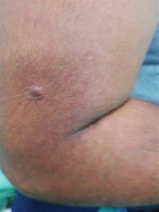
Case presentation: We report a rare case of a 38-year-old man who presented with a painful nodule on his right upper arm. A definite diagnosis was made by histopathological study. A complete surgical excision was performed to avoid recurrence.
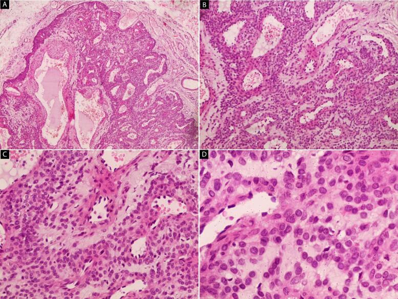
Clinical discussion: Glomus tumors form less than 2 % of all soft tissue tumors. The tumor was first reported by Wood in 1812. It typically appears like a small blue-red solitary papule in the hand especially the digits, which are the most prevalent location for glomus tumors with an incidence rate of up to 75 %. The histopathology findings of glomus tumor, are three components: glomus cells, vasculature, and smooth muscle cells. The preferred method of treatment is total excision to prevent a recurrence.
Conclusion: Eventually, the glomus tumor is fairly a rare benign tumor that physicians should keep in mind as a deferential diagnosis when facing a subcutaneous nodule and don't rule out when the tumor is extradigital.
Keywords: Benign; Extradigital; Glomus tumor; Nodule; Surgery; Upper arm.
Copyright © 2024 The Authors. Published by Elsevier Ltd.. All rights reserved.
Conflict of interest statement
Conflict of interest statement The authors have no conflicts of interest to declare.
Figures
Similar articles
-
Extradigital glomus tumor: A case report.Mol Clin Oncol. 2014 Mar;2(2):237-239. doi: 10.3892/mco.2013.219. Epub 2013 Dec 4. Mol Clin Oncol. 2014. PMID: 24649339 Free PMC article.
-
Extradigital Glomus Tumor Revisited: Painful Subcutaneous Nodules Located in Various Parts of the Body.Indian J Dermatol. 2016 Jan-Feb;61(1):118. doi: 10.4103/0019-5154.174080. Indian J Dermatol. 2016. PMID: 26955123 Free PMC article.
-
Glomus Extradigital Tumor: A Case Report of an Extradigital Glomus Tumor on the Wrist and Comprehensive Review of Glomus Tumors.Cureus. 2023 May 8;15(5):e38737. doi: 10.7759/cureus.38737. eCollection 2023 May. Cureus. 2023. PMID: 37292537 Free PMC article.
-
Large solitary glomus tumor of the wrist involving the radial artery.Am J Orthop (Belle Mead NJ). 2014 Dec;43(12):567-70. Am J Orthop (Belle Mead NJ). 2014. PMID: 25490012 Review.
-
Extradigital Symplastic Glomus Tumor of the Hand: Report of 2 Cases and Literature Review.Am J Dermatopathol. 2015 Jul;37(7):560-2. doi: 10.1097/DAD.0000000000000132. Am J Dermatopathol. 2015. PMID: 25051107 Review.
Cited by
-
Diagnosis and Excision of Glomangioma of the Lower Extremity.Cureus. 2025 Mar 12;17(3):e80458. doi: 10.7759/cureus.80458. eCollection 2025 Mar. Cureus. 2025. PMID: 40225461 Free PMC article.
References
-
- Hrubý J., Novotný R., Spaček M., Mitáš P., Hlubocký J., Janák D., Povýšil C., Lindner J. Surgical extirpation of glomus tumor from rare localization on the upper extremity. Case Rep. Vasc. Med. 2013;2013 doi: 10.1155/2013/570945. (Epub 2013 Sep 25. PMID: 24187644; PMCID: PMC3800625) - DOI - PMC - PubMed
Publication types
LinkOut - more resources
Full Text Sources
